Chapter Preamble
Signals declared
Sig-D — Diagnostic (primary). Diagnose ADPKD by the ultrasound criteria, MRI, or genetics; distinguish it from other cystic diseases; and recognise its manifestations.
Sig-T — Therapeutic (strong). Risk-stratify to identify rapid progressors, and manage the renal manifestations — hypertension, pain, haematuria, cyst infection, stones.
Sig-V — Evidence-dense (strong). The Mayo imaging classification and the PROPKD score as validated risk-prediction tools — graded and reflected on.
Levels populated and omitted
Populated (18): L1–L5, L7, L8, L10–L14, L17–L22. The evidence and therapeutic signals fire the absolute-risk table (L14), the templates (L17), and the reflective prompts (L21); the diagnostic signal drives the tables, rules, cases, pitfalls, and board items.
L6 / L9 mechanism levels — omitted. No Sig-M; the mechanism was built in Chapter 4, and this is a diagnosis, risk, and management chapter.
L15 / L16 preference-sensitive map and SDM scripts — omitted. No Sig-E; diagnosis and risk-stratification are evidence-driven, with the preference-sensitive aspects of tolvaptan in the next chapter.
| 01 | PHASE A · LEVEL 1 · ORIENTATION & KNOWLEDGE Learning Objectives |
By the end of this chapter you should be able to:
Apply the ultrasound diagnostic criteria for ADPKD in at-risk individuals.
Decide when MRI or genetic testing is needed for diagnosis or exclusion.
Distinguish ADPKD from other cystic kidney diseases.
Risk-stratify with the Mayo imaging classification and the PROPKD score.
Explain the purpose of risk-stratification — identifying rapid progressors.
Recognise and manage the renal manifestations of ADPKD.
Distinguish cyst infection from cyst haemorrhage and treat each.
Recognise nephrolithiasis and the systemic manifestations.
| 02 | PHASE A · LEVEL 2 · ORIENTATION & KNOWLEDGE Executive Summary |
ADPKD is diagnosed in at-risk individuals by age-based ultrasound criteria — more cysts being required at older ages — with specific criteria allowing exclusion (important for donor evaluation).
MRI or genetic testing is used when ultrasound is insufficient — in the young at-risk, in potential living donors, with no family history, or in atypical cases.
About one in eight patients has no family history (a de novo mutation), where genetic testing helps.
ADPKD must be distinguished from other cystic diseases — ARPKD, ADTKD, acquired cystic disease, simple cysts, and the cystic disease of tuberous sclerosis and von Hippel-Lindau.
Risk-stratification identifies the rapid progressors who benefit from disease-modifying treatment.
The Mayo imaging classification combines height-adjusted total kidney volume with age to assign a risk class, with the higher classes marking rapid progressors.
The PROPKD score combines clinical and genetic factors — sex, early hypertension, early urological events, and genotype — to predict the age at end-stage disease.
Hypertension is an early and common renal manifestation, treated with RAAS blockade.
Pain is common — chronic from the enlarging kidneys, acute from cyst haemorrhage, infection, or stones.
Haematuria from cyst haemorrhage is common and usually self-limiting.
Cyst infection is hard to diagnose — fever and localised pain, often with a negative urine culture — and is treated with lipophilic antibiotics that penetrate cysts, such as fluoroquinolones.
Nephrolithiasis is more common than in the general population, with uric acid and calcium oxalate stones, from urinary stasis and low citrate.
The systemic manifestations — liver cysts, intracranial aneurysms, and others — complete the picture, and recognising and managing all of these is the substance of ADPKD care.
| 03 | PHASE A · LEVEL 3 · ORIENTATION & KNOWLEDGE Main Narrative |
With the genetics and mechanism of ADPKD established, this chapter is about the patient in front of you: making the diagnosis, predicting how fast the disease will progress, and recognising and managing its many manifestations. Diagnosis is increasingly nuanced — ultrasound for most, but MRI or genetics where it falls short; risk prediction has become central, because identifying rapid progressors selects who benefits from treatment; and the manifestations, from hypertension to cyst infection, are the day-to-day work of ADPKD care.
— Diagnosis: ultrasound, MRI, genetics
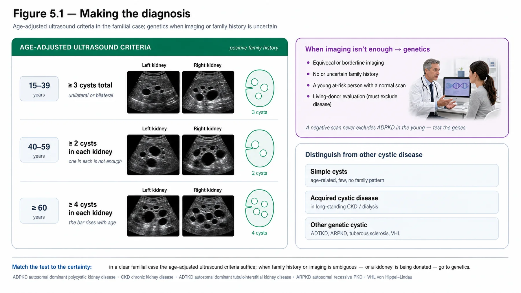
ADPKD is most often diagnosed by ultrasound in an at-risk individual (someone with an affected first-degree relative), using validated age-based criteria: because cysts accumulate with age, more cysts are required to make the diagnosis at older ages — broadly, a small number of cysts suffices in a young adult, more in middle age, and more again in older age. Equally important are the exclusion criteria, which allow ADPKD to be ruled out in an older at-risk relative — critical when evaluating a potential living kidney donor. Ultrasound's limitation, met in the imaging chapter, is its insensitivity for the few small cysts of a young at-risk person, so MRI (more sensitive) or genetic testing is used when ultrasound is insufficient: in the young at-risk, in potential donors (where the disease must be definitively excluded), where the family history is absent or unknown, and in atypical presentations. Note that roughly one in eight patients has no family history — a de novo mutation — so the absence of a family history does not exclude ADPKD, and genetic testing is particularly useful there. Genetic testing also has a role in reproductive planning and in establishing the gene for prognosis. So the diagnostic approach is ultrasound first, MRI or genetics where it falls short, and a low threshold for genetics in the young, the donor, and the atypical.
— The differential of cystic kidneys
Not every cystic kidney is ADPKD, and distinguishing the alternatives matters. ARPKD (autosomal recessive polycystic kidney disease) presents in infancy or childhood, is recessive (affected siblings, unaffected parents), and has a different appearance and associated congenital hepatic fibrosis (the next chapter). ADTKD (autosomal dominant tubulointerstitial kidney disease) is dominant like ADPKD but has few or no cysts and a tubulointerstitial, bland-urine picture (a later chapter). Acquired cystic kidney disease develops in long-standing CKD and dialysis patients (not inherited, with a risk of malignancy). Simple cysts are common, age-related, and few — not ADPKD. And the cystic disease of the phakomatoses must be considered: tuberous sclerosis (cysts with angiomyolipomas) and von Hippel-Lindau (cysts with a risk of clear-cell renal cell carcinoma), both with their characteristic extrarenal features. Distinguishing these rests on the age and mode of onset, the family history and inheritance pattern, the number and character of the cysts, the associated features (hepatic fibrosis, angiomyolipomas, tumours), and, increasingly, genetics. ADPKD is the commonest, but the differential must be kept in mind, especially when the picture is atypical.
— Risk prediction: who progresses fast
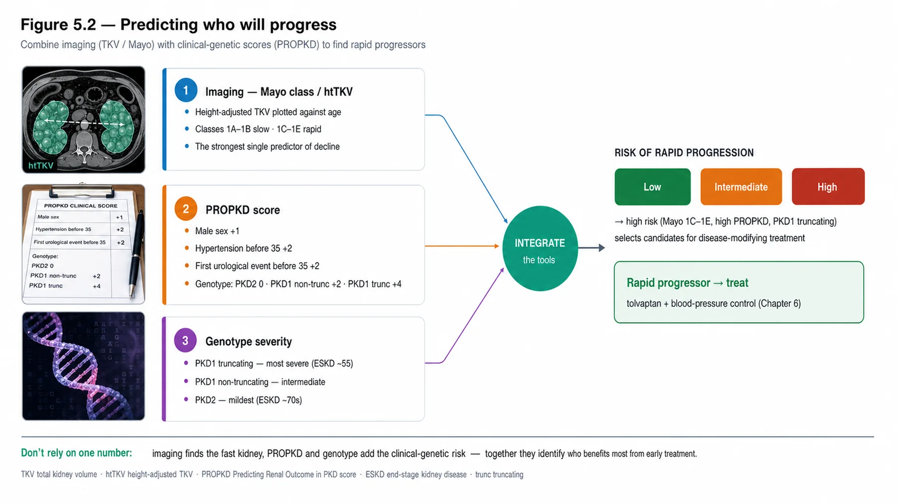
A defining advance in ADPKD care is risk prediction — identifying, before the GFR falls, which patients will progress rapidly, because they are the ones who benefit most from disease-modifying treatment. Two validated tools dominate. The Mayo imaging classification uses the height-adjusted total kidney volume together with age to assign a patient to a risk class: it captures the cyst burden relative to age (a large kidney volume at a young age predicts rapid progression), and the higher classes mark the rapid progressors who are candidates for treatment. The PROPKD score takes a complementary, clinical-and-genetic approach, scoring points for male sex, hypertension before age 35, the first urological event (haematuria, pain, infection) before age 35, and the genotype (PKD2 lowest risk, PKD1 non-truncating intermediate, PKD1 truncating highest) — stratifying patients into low, intermediate, and high risk and predicting the age at end-stage disease. Together with the rate of GFR decline and the family history of early kidney failure, these tools turn ADPKD from a disease with a vague 'you'll need dialysis someday' prognosis into one where the individual's trajectory can be estimated and the rapid progressors identified for treatment. Risk-stratification is the bridge between diagnosis and the treatment decision of the next chapter.
— Hypertension, pain, and haematuria
The renal manifestations of ADPKD are the substance of its day-to-day management. Hypertension is early and common — appearing often before the GFR falls, driven by the activation of the renin-angiotensin system as expanding cysts compress the intrarenal vasculature — and it both reflects and accelerates the disease, so it is treated, with RAAS blockade the preferred first-line agent (renoprotective; the next chapter develops the targets). Pain is one of the commonest complaints: chronic flank or abdominal pain from the mass effect of the enlarging kidneys and liver, and acute pain from a specific event — cyst haemorrhage, cyst infection, a stone, or (rarely) cyst rupture. Distinguishing the cause of acute pain is the key clinical task, because the management differs. Haematuria is common and usually due to cyst haemorrhage (bleeding into a cyst, which may communicate with the collecting system); it is typically self-limiting, managed conservatively with rest and hydration, though it can recur. These three — hypertension to be treated, pain to be characterised by cause, and haematuria usually to be managed conservatively — are the everyday face of ADPKD.
— Cyst infection, stones, and the systemic disease
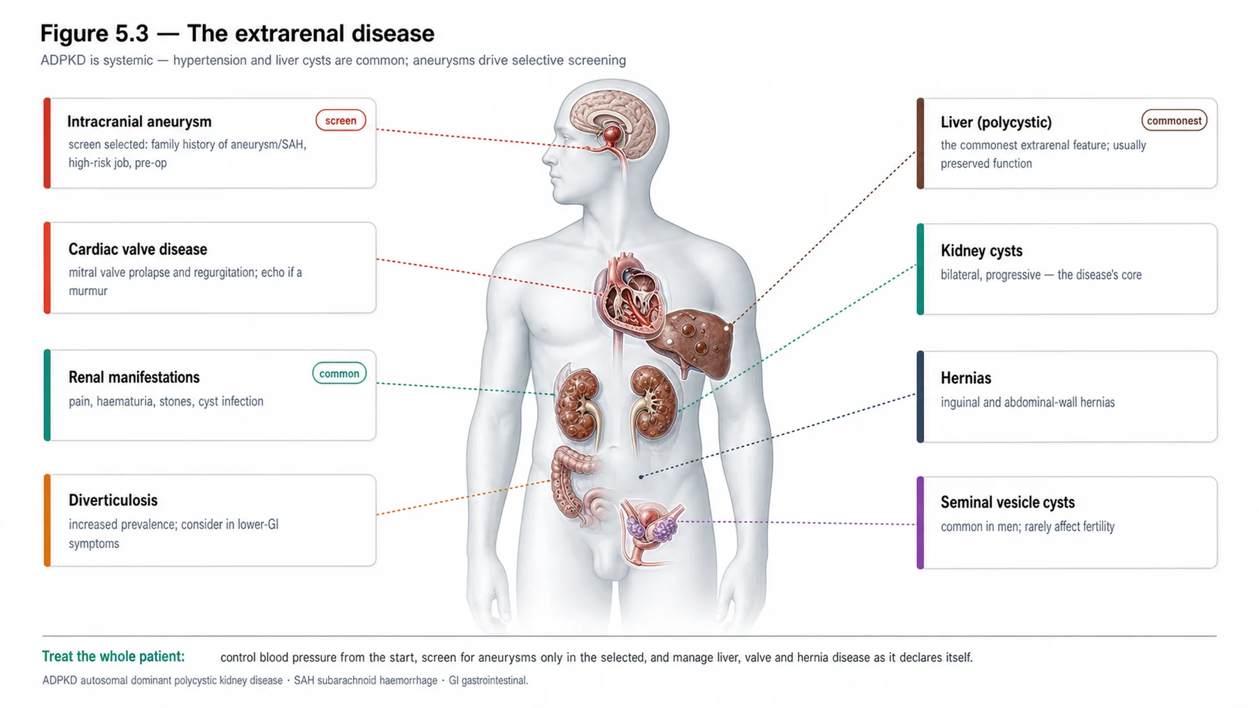
Two manifestations deserve particular attention. Cyst infection is notoriously difficult to diagnose and treat: it presents with fever and localised flank pain, but the urine culture is often negative (because the infected cyst may not communicate with the urine), so the diagnosis rests on the clinical picture and imaging (CT or MRI, and sometimes PET, which can localise an infected cyst). Crucially, it must be treated with lipophilic antibiotics that penetrate the cyst wall — fluoroquinolones and co-trimoxazole are the classic choices, whereas many hydrophilic antibiotics fail to enter cysts — often for a prolonged course, with drainage of a large or refractory infected cyst when antibiotics alone fail. Recognising that an ADPKD patient with fever and flank pain may have a cyst infection requiring cyst-penetrating antibiotics (not a standard urinary-tract-infection regimen) is an important practical point. Nephrolithiasis is also more common in ADPKD (around a fifth of patients), with uric acid and calcium oxalate stones favoured by urinary stasis (from the distorted anatomy), a low urine citrate, and a low urine pH; it is managed as stones are generally, with attention to these factors (the stone chapter). And the systemic manifestations of the previous chapter — liver cysts, intracranial aneurysms, valve disease — round out the picture. Recognising and managing this full range of manifestations, on a foundation of diagnosis and risk-stratification, is what comprehensive ADPKD care entails — and the next chapter turns to the disease-modifying treatment that risk-stratification selects for.
| 04 | PHASE A · LEVEL 4 · ORIENTATION & KNOWLEDGE Reference Tables |
Table 5.1 — Diagnosis of ADPKD
| Modality | Use |
| Ultrasound | First-line in at-risk individuals — age-based cyst criteria; exclusion criteria for donors |
| MRI | More sensitive — young at-risk, donor evaluation, atypical |
| Genetic testing | No/unknown family history, donor, reproductive planning, atypical; establishes the gene |
| No family history | ~1 in 8 (de novo) — absence does not exclude ADPKD |
Table 5.2 — Differential of cystic kidneys
| Condition | Distinguishing feature |
| ARPKD | Infantile/childhood, recessive, congenital hepatic fibrosis |
| ADTKD | Dominant but few/no cysts; tubulointerstitial, bland urine |
| Acquired cystic / simple cysts | In CKD/dialysis (malignancy risk); or age-related, few |
| Tuberous sclerosis / VHL | Cysts + angiomyolipomas (TSC); cysts + clear-cell RCC (VHL) |
Table 5.3 — Risk prediction
| Tool | Detail |
| Mayo imaging classification | Height-adjusted TKV + age → risk class; higher classes = rapid progressors |
| PROPKD score | Male sex, HTN < 35, urological event < 35, genotype → low/intermediate/high risk |
| Genotype within PROPKD | PKD2 < PKD1 non-truncating < PKD1 truncating (rising risk) |
| Purpose | Identify rapid progressors who benefit from disease-modifying treatment |
Table 5.4 — Renal manifestations
| Manifestation | Detail |
| Hypertension | Early, common (RAAS activation); treat (RAAS blockade first-line) |
| Pain | Chronic (mass effect); acute (haemorrhage, infection, stone, rupture) |
| Haematuria | Cyst haemorrhage — common, usually self-limiting (rest, hydration) |
| Stones / concentrating defect | Nephrolithiasis (~20%); reduced urinary concentrating ability (early) |
Table 5.5 — Cyst infection versus cyst haemorrhage
| Feature | Cyst infection | Cyst haemorrhage |
| Presentation | Fever, localised pain | Pain ± haematuria, no fever |
| Diagnosis | Often negative urine culture; imaging (CT/MRI/PET) | Clinical ± imaging |
| Treatment | Cyst-penetrating antibiotics (fluoroquinolone, co-trimoxazole) ± drainage | Conservative (rest, hydration) |
Table 5.6 — Stones and the systemic disease
| Aspect | Detail |
| Nephrolithiasis | ~20% — uric acid and calcium oxalate; stasis, low citrate, low pH |
| Stone management | As usual (Chapter 16) — hydration, citrate, address the factors |
| Systemic (Ch 4) | Liver cysts (commonest), intracranial aneurysms, valves, diverticulosis, hernias |
| Comprehensive care | Diagnosis + risk-stratification + manifestation management + systemic surveillance |
| 05 | PHASE B · LEVEL 5 · VISUALISE & MAP Imaging & Flowchart Specifications |
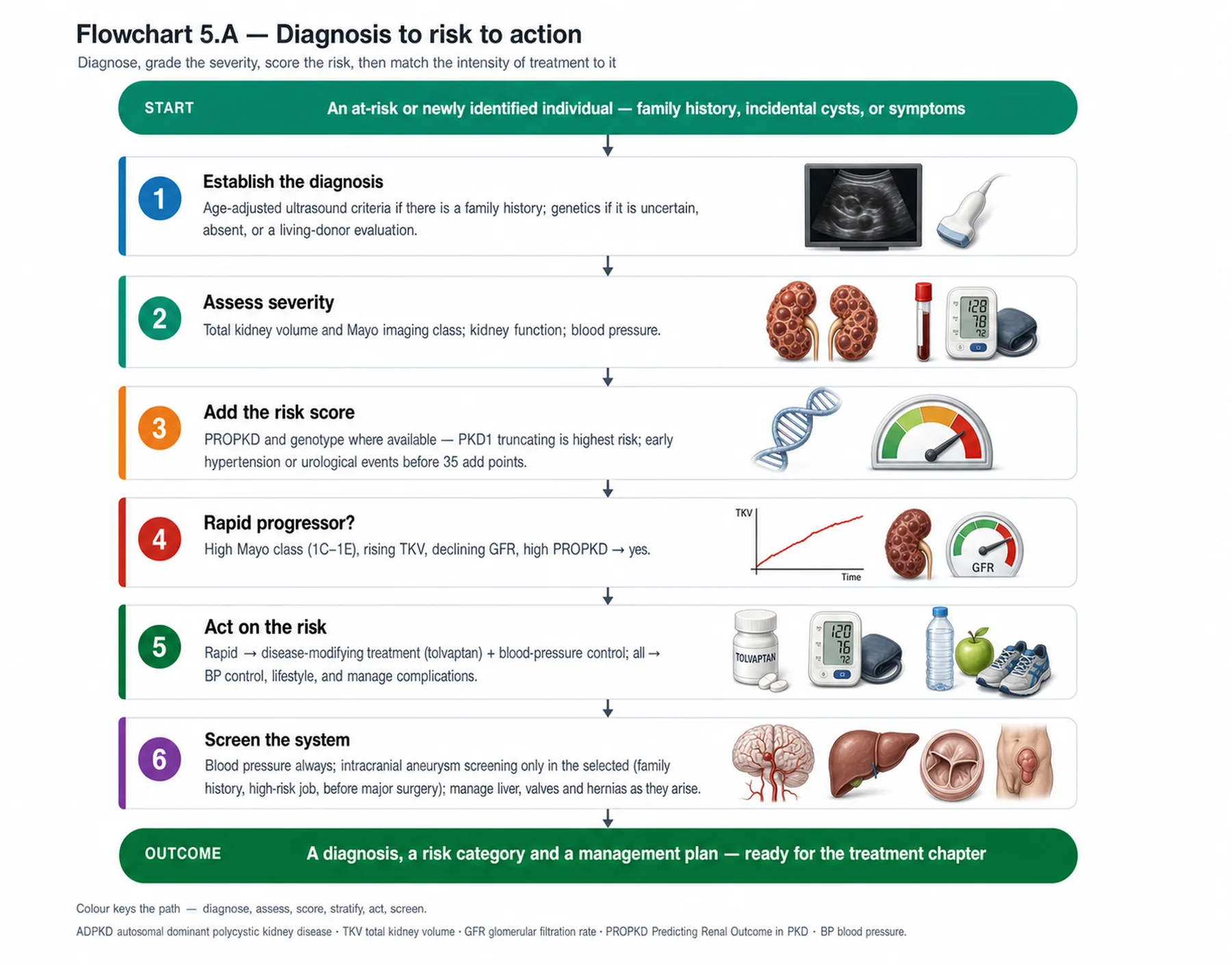
| 08 | PHASE C · LEVEL 8 · CLINICAL REASONING Clinical Cases |
| CASE 1 | MAKE — OR EXCLUDE — THE DIAGNOSIS Criteria, MRI, genetics Diagnosing ADPKD |
Presentation
A young at-risk adult with a few cysts on ultrasound, and a separate older at-risk relative being assessed as a potential donor, both need a definitive answer about whether they have ADPKD. The team applies a single rule of thumb to both.
❖ Pause and reflect How should the diagnosis be made or excluded in each? |
Analysis
The age-based criteria mean the two need different handling. ADPKD is diagnosed in at-risk individuals by ultrasound criteria that require more cysts at older ages (because cysts accumulate), and there are separate exclusion criteria for ruling it out. For the young at-risk adult with only a few cysts, ultrasound may be inconclusive — a small number of cysts can be diagnostic at a young age, but ultrasound is also insensitive for early disease — so MRI (more sensitive) or genetic testing may be needed to be definitive. For the older at-risk relative being assessed as a donor, the priority is to definitively exclude ADPKD before donation: the exclusion criteria (or genetics/MRI) must rule it out, because transplanting an affected kidney would harm both. A single rule of thumb ignores the age-dependence of the criteria and the donor's need for definitive exclusion. Each needs the appropriate test.
Plan
Apply the age-based ultrasound criteria, using MRI or genetics for the young at-risk adult (definitive diagnosis) and to definitively exclude ADPKD in the older potential donor before donation. Use the age-appropriate criteria and a low threshold for MRI/genetics in the young and the donor.
Teaching point
ADPKD ultrasound criteria are age-based; use MRI or genetics when ultrasound is insufficient — in the young at-risk and, critically, to exclude the disease in a potential donor.
Cross-reference
Exercises the diagnosis content; Figure 5.1; Tables 5.1, 5.2; imaging in Chapter 3; donor evaluation in Chapter 1.
| CASE 2 | WHO PROGRESSES FAST? Mayo and PROPKD Risk prediction |
Presentation
Two ADPKD patients with similar current eGFRs are being managed identically, with no risk-stratification, and the team cannot say which one is likely to progress quickly or who might benefit from disease-modifying treatment.
❖ Pause and reflect How can rapid progressors be identified before the GFR falls? |
Analysis
With the validated risk-prediction tools — the Mayo imaging classification and the PROPKD score — which identify rapid progressors before the GFR declines. The Mayo classification combines the height-adjusted total kidney volume with age to assign a risk class, with the higher classes marking rapid progressors (a large kidney volume for the patient's age predicts fast progression even when the current eGFR is similar). The PROPKD score adds clinical and genetic factors — male sex, hypertension before 35, an early urological event, and the genotype — to stratify risk and predict the age at end-stage disease. Two patients with the same current eGFR can have very different trajectories, and these tools reveal it. Risk-stratification is not academic: it identifies who is likely to progress rapidly and therefore who stands to benefit most from disease-modifying treatment (the next chapter), so managing both patients identically without stratifying misses this.
Plan
Risk-stratify both patients with the Mayo imaging classification (height-adjusted TKV + age) and the PROPKD score (clinical + genetic) to identify the rapid progressor and inform the treatment decision, rather than managing identically on the current eGFR. Risk-stratify to find the rapid progressors.
Teaching point
Use the Mayo imaging classification and the PROPKD score to identify rapid progressors before the GFR falls — they select who benefits from disease-modifying treatment.
Cross-reference
Exercises the risk-prediction content; Figure 5.2; Table 5.3; TKV in Chapter 3; tolvaptan in Chapter 6.
| CASE 3 | FEVER AND FLANK PAIN Cyst-penetrating antibiotics Cyst infection |
Presentation
An ADPKD patient presents with fever and localised flank pain, but the urine culture is negative. A clinician, seeing the negative culture, doubts infection and considers stopping antibiotics.
❖ Pause and reflect Does a negative urine culture exclude a cyst infection? |
Analysis
No — a negative urine culture does not exclude a cyst infection, which is notoriously difficult to diagnose. The infected cyst often does not communicate with the collecting system, so the urine culture is frequently negative despite an active cyst infection; the diagnosis rests on the clinical picture (fever and localised flank pain) and imaging (CT or MRI, and sometimes PET, which can localise an infected cyst). Crucially, cyst infection must be treated with lipophilic antibiotics that penetrate the cyst wall — fluoroquinolones or co-trimoxazole — because many hydrophilic antibiotics fail to enter the cyst, and often for a prolonged course, with drainage of a large or refractory infected cyst if antibiotics alone fail. Stopping antibiotics on the negative culture, or using a standard urinary-tract-infection regimen that does not penetrate cysts, would undertreat a serious infection. The negative culture is expected, not reassuring.
Plan
Treat the likely cyst infection despite the negative urine culture, using cyst-penetrating antibiotics (a fluoroquinolone or co-trimoxazole) for a prolonged course, with imaging to localise and drainage if refractory. A negative culture does not exclude cyst infection — use cyst-penetrating antibiotics.
Teaching point
Cyst infection often has a negative urine culture — diagnose clinically and by imaging, and treat with cyst-penetrating antibiotics (fluoroquinolone, co-trimoxazole), not a standard UTI regimen.
Cross-reference
Exercises the cyst-infection content; Figure 5.3; Tables 5.4, 5.5.
| CASE 4 | PAIN, BLOOD, AND STONES Managing the manifestations Renal manifestations |
Presentation
An ADPKD patient has chronic flank pain, an episode of gross haematuria, and a history of kidney stones, and the team is unsure how to approach each.
❖ Pause and reflect How should each of these manifestations be managed? |
Analysis
Each manifestation has a characteristic approach. The chronic flank pain is most likely from the mass effect of the enlarging kidneys (and liver), managed conservatively in a stepwise way (lifestyle, analgesia, and interventional options for refractory pain), after excluding an acute cause. The gross haematuria is most likely from cyst haemorrhage — common and usually self-limiting — managed conservatively with rest and hydration, with reassurance that it typically settles (though it can recur). The kidney stones reflect the increased nephrolithiasis of ADPKD (around a fifth of patients), with uric acid and calcium oxalate stones favoured by urinary stasis, low urine citrate, and low pH; they are managed as stones generally are (hydration, citrate, addressing the metabolic factors). Recognising each manifestation's usual cause and management — conservative for chronic pain and haematuria, stone management with attention to the ADPKD-specific factors — is the everyday work of ADPKD care, alongside treating the hypertension and watching for the acute complications.
Plan
Manage the chronic pain conservatively and stepwise (after excluding an acute cause), the cyst-haemorrhage haematuria conservatively (rest, hydration), and the stones with hydration, citrate, and attention to the ADPKD-specific factors. Manage each manifestation by its usual cause and approach.
Teaching point
ADPKD manifestations have characteristic management — chronic pain and cyst-haemorrhage haematuria conservatively, stones with hydration/citrate — alongside treating the hypertension and recognising acute complications.
Cross-reference
Exercises the manifestations content; Tables 5.4, 5.6; stones in Chapter 16.
| 10 | PHASE C · LEVEL 10 · CLINICAL REASONING Clinical Pearls |
| Diagnose ADPKD in at-risk individuals by age-based ultrasound criteria (more cysts at older ages). | Exclusion criteria allow ruling it out — critical for donor evaluation. |
| Use MRI or genetics when ultrasound is insufficient (young, donor, no family history, atypical). | ~1 in 8 have no family history (de novo) — absence doesn't exclude ADPKD. |
| Differential: ARPKD, ADTKD, acquired cystic, simple cysts, tuberous sclerosis, VHL. | Risk-stratify to identify rapid progressors (who benefit from treatment). |
| Mayo classification: height-adjusted TKV + age → risk class (higher = rapid). | PROPKD score: sex, HTN < 35, urological event < 35, genotype → low/intermediate/high. |
| Genotype risk: PKD2 < PKD1 non-truncating < PKD1 truncating. | Hypertension is early and common — treat (RAAS blockade first-line). |
| Pain: chronic (mass effect) vs acute (haemorrhage, infection, stone, rupture). | Haematuria (cyst haemorrhage) — common, usually self-limiting (rest, hydration). |
| Cyst infection: fever + localised pain, often NEGATIVE urine culture. | Treat cyst infection with cyst-penetrating antibiotics (fluoroquinolone, co-trimoxazole). |
| Nephrolithiasis ~20% (uric acid, calcium oxalate; stasis, low citrate). | Systemic: liver cysts, aneurysms, valves — part of comprehensive care. |
| 11 | PHASE D · LEVEL 11 · SAFETY & EVIDENCE Red Flags & Never-Do |
Panel A — Red flags
| ▲ | A young at-risk person or potential donor needing definitive diagnosis/exclusion — ultrasound may not suffice; use MRI/genetics. |
| ▲ | No risk-stratification in an ADPKD patient — the rapid progressor (and treatment candidate) is unidentified. |
| ▲ | Fever and flank pain with a negative urine culture — suspect cyst infection; use cyst-penetrating antibiotics. |
| ▲ | Early hypertension in ADPKD — treat it (RAAS blockade first-line). |
| ▲ | Acute flank pain in ADPKD — characterise the cause (haemorrhage, infection, stone, rupture). |
Panel B — Never do
| ✖ NEVER — exclude ADPKD on a normal ultrasound in the young or a donor without MRI/genetics. |
| ✖ NEVER — manage every ADPKD patient identically without risk-stratification. |
| ✖ NEVER — exclude cyst infection on a negative urine culture. |
| ✖ NEVER — treat cyst infection with a non-penetrating standard UTI regimen. |
| 12 | PHASE D · LEVEL 12 · SAFETY & EVIDENCE Common Pitfalls |
Pitfall 1 — One rule for all ages
| ✖ | WRONG Applying a single cyst-count rule regardless of age. |
| ✓ | RIGHT Using the age-based criteria and MRI/genetics where needed. |
| ✉ | WHY Cyst counts required differ by age; ultrasound is insensitive in the young. |
Pitfall 2 — No risk-stratification
| ✖ | WRONG Managing ADPKD patients identically on current eGFR. |
| ✓ | RIGHT Risk-stratifying with the Mayo classification and PROPKD score. |
| ✉ | WHY These identify rapid progressors before the GFR falls. |
Pitfall 3 — Trusting the negative culture
| ✖ | WRONG Excluding cyst infection because the urine culture is negative. |
| ✓ | RIGHT Treating clinically with cyst-penetrating antibiotics. |
| ✉ | WHY Infected cysts often don't communicate with the urine. |
Pitfall 4 — Wrong antibiotic
| ✖ | WRONG Using a standard UTI antibiotic for a cyst infection. |
| ✓ | RIGHT Using a lipophilic, cyst-penetrating antibiotic (fluoroquinolone, co-trimoxazole). |
| ✉ | WHY Many antibiotics fail to penetrate the cyst wall. |
Pitfall 5 — Missing the acute cause of pain
| ✖ | WRONG Treating acute flank pain as chronic mass-effect pain. |
| ✓ | RIGHT Characterising the acute cause (haemorrhage, infection, stone, rupture). |
| ✉ | WHY Acute causes need specific management. |
| 13 | PHASE D · LEVEL 13 · SAFETY & EVIDENCE Evidence Grading |
GRADE A HIGH CONFIDENCE The effect is real and the estimate is stable. RCTs at low risk of bias; multiple concordant prospective cohorts; meta-analyses. |
GRADE B MODERATE CONFIDENCE The effect is likely real but may shift with new data. Observational studies, registries, mechanistic human studies. |
GRADE C LOW CONFIDENCE Rests on physiology, reasoning, or consensus rather than outcomes. Pathophysiological reasoning; extrapolation; consensus without outcomes. |
Graded statements (by evidence type)
| Statement | Grade | Basis (evidence type) |
| Age-based ultrasound criteria diagnose ADPKD in at-risk individuals. | A | Validation studies |
| The Mayo imaging classification predicts progression. | A | Validation against outcomes |
| The PROPKD score predicts the age at end-stage disease. | A | Validation cohorts |
| Hypertension is early and common and should be treated. | A | Clinical and trial data |
| Cyst infection often has a negative urine culture and needs penetrating antibiotics. | B | Clinical data and pharmacology |
| Nephrolithiasis is more common in ADPKD. | A | Epidemiological data |
| Risk-stratification identifies candidates for disease-modifying treatment. | A | Guidelines and trial selection |
| 14 | PHASE E · LEVEL 14 · PATIENT DECISIONS Absolute Risk in Natural Frequency |
Natural-frequency estimates for orientation, from ADPKD cohorts; they vary with the population. They convey the size of the decisions, expressed per 100 comparable patients.
| Per 100 patients… | Outcome | Roughly how many | See |
| ADPKD patients | Have nephrolithiasis | Around 20 — more than the general population | L13 row 6 |
| ADPKD patients | Have no family history (de novo) | Around 12 — hence absence doesn't exclude | L13 row 1 |
| Risk-stratified by Mayo/PROPKD | Are identified as rapid progressors | A meaningful subset — treatment candidates | L13 rows 2–3 |
| With cyst infection | Have a negative urine culture | Many — hence don't exclude on culture | L13 row 5 |
★ How to read these Read these as orientation, not promises; proportions vary with the population. The stable signals: stones are common, a meaningful minority have no family history, risk tools identify the rapid progressors who benefit from treatment, and cyst infection often has a negative culture. Communicate them as people out of 100, not as a hazard ratio. |
| 17 | PHASE F · LEVEL 17 · APPLY & TEST Documentation Templates |
Paste-ready notes. Tick the boxes that apply and delete the rest; make the diagnosis, the risk class, and the manifestation plan explicit.
Template 1 — Diagnosis and risk-stratification
Template 2 — Manifestation management
| 18 | PHASE F · LEVEL 18 · APPLY & TEST Cheat Sheet |
| Diagnose by age-based ultrasound criteria (more cysts at older ages). | Exclusion criteria — critical for donors. |
| MRI/genetics if young, donor, no family history, atypical. | ~1 in 8 de novo — no family history doesn't exclude. |
| Differential: ARPKD, ADTKD, acquired, simple, TSC, VHL. | Risk-stratify to find rapid progressors. |
| Mayo: htTKV + age → risk class. | PROPKD: sex, HTN<35, urological event<35, genotype. |
| Genotype risk: PKD2 < PKD1 non-truncating < PKD1 truncating. | Hypertension early/common — treat (RAAS blockade). |
| Pain: chronic (mass) vs acute (haemorrhage/infection/stone/rupture). | Haematuria (cyst haemorrhage) — conservative. |
| Cyst infection: fever + pain, often NEGATIVE culture. | Cyst infection → cyst-penetrating antibiotics (fluoroquinolone/co-trimoxazole). |
| Stones ~20% (uric acid, calcium oxalate). | Systemic: liver cysts, aneurysms, valves. |
| 19 | PHASE F · LEVEL 19 · APPLY & TEST Flashcards |
| CARD 1 | Q. How is ADPKD diagnosed in an at-risk individual? A. By age-based ultrasound criteria — requiring more cysts at older ages because cysts accumulate — with separate exclusion criteria for ruling it out; MRI or genetic testing is used when ultrasound is insufficient, as in the young at-risk, a potential donor, no family history, or atypical cases. DETAILED. About one in eight have no family history (de novo). CLINICAL. Use age-appropriate criteria, with MRI/genetics in the young and the donor. |
| CARD 2 | Q. What must ADPKD be distinguished from? A. Other cystic kidney diseases — ARPKD (infantile, recessive, hepatic fibrosis), ADTKD (dominant but few/no cysts, tubulointerstitial), acquired cystic disease (in CKD/dialysis), simple cysts (age-related, few), and the cystic disease of tuberous sclerosis (with angiomyolipomas) and von Hippel-Lindau (with clear-cell carcinoma). DETAILED. Age, inheritance, cyst character, and associated features distinguish them. CLINICAL. Keep the cystic-disease differential in mind, especially when atypical. |
| CARD 3 | Q. What are the two main risk-prediction tools in ADPKD? A. The Mayo imaging classification (height-adjusted total kidney volume plus age, assigning a risk class with the higher classes marking rapid progressors) and the PROPKD score (male sex, hypertension before 35, an early urological event, and genotype, stratifying low/intermediate/high risk and predicting the age at end-stage disease). DETAILED. They identify rapid progressors before the GFR falls. CLINICAL. Risk-stratify with Mayo and PROPKD to select treatment candidates. |
| CARD 4 | Q. Why and how is hypertension managed in ADPKD? A. Because it is early, common (from renin-angiotensin activation as cysts compress the vasculature), and both reflects and accelerates the disease; it is treated, with RAAS blockade the preferred first-line agent for its renoprotection. DETAILED. It often appears before the GFR falls. CLINICAL. Treat the hypertension with RAAS blockade. |
| CARD 5 | Q. How does cyst infection present and how is it treated? A. With fever and localised flank pain, often with a negative urine culture (because the infected cyst may not communicate with the urine); diagnosed clinically and by imaging (CT/MRI, sometimes PET), and treated with lipophilic, cyst-penetrating antibiotics (fluoroquinolones or co-trimoxazole) for a prolonged course, with drainage if refractory. DETAILED. A negative culture does not exclude it. CLINICAL. Treat with cyst-penetrating antibiotics despite a negative culture. |
| CARD 6 | Q. How is haematuria in ADPKD usually managed? A. It is usually due to cyst haemorrhage (bleeding into a cyst), is common, and is typically self-limiting — managed conservatively with rest and hydration — though it can recur. DETAILED. It is generally benign. CLINICAL. Manage cyst-haemorrhage haematuria conservatively. |
| CARD 7 | Q. Why is nephrolithiasis more common in ADPKD? A. Because the distorted anatomy causes urinary stasis, and there is often a low urine citrate and low urine pH, favouring uric acid and calcium oxalate stones; it affects around a fifth of patients and is managed with hydration, citrate, and attention to these factors. DETAILED. The stones reflect ADPKD-specific factors. CLINICAL. Manage stones with hydration and citrate, addressing the factors. |
| CARD 8 | Q. What is the purpose of risk-stratification in ADPKD? A. To identify, before the GFR falls, which patients will progress rapidly — because they are the ones who benefit most from disease-modifying treatment (the next chapter) — turning a vague prognosis into an individualised trajectory. DETAILED. It bridges diagnosis and the treatment decision. CLINICAL. Risk-stratify to select who benefits from treatment. |
| 20 | PHASE F · LEVEL 20 · APPLY & TEST One-Minute Preceptor |
| SCENE 1 | The intern doubting cyst infection |
GET A COMMITMENT. “This ADPKD patient has fever and flank pain but a negative urine culture — you don't think it's infection?”
PROBE FOR EVIDENCE. “The culture is negative” — ask: “Why might an infected cyst give a negative urine culture, and what antibiotics reach a cyst?”
TEACH A GENERAL RULE. An infected cyst often doesn't communicate with the urine, so the culture is negative; diagnose clinically and by imaging, and treat with cyst-penetrating antibiotics (fluoroquinolone, co-trimoxazole).
REINFORCE WHAT WAS RIGHT. Checking the culture was reasonable.
CORRECT A MISTAKE. Treat the cyst infection despite the negative culture, with penetrating antibiotics.
| SCENE 2 | The resident skipping risk-stratification |
GET A COMMITMENT. “You're managing these two ADPKD patients identically on their eGFR — can you say who'll progress faster?”
PROBE FOR EVIDENCE. “Their eGFRs are similar” — ask: “What tools predict progression before the GFR falls, and why does it matter?”
TEACH A GENERAL RULE. The Mayo imaging classification and PROPKD score identify rapid progressors before the GFR declines — selecting who benefits from disease-modifying treatment.
REINFORCE WHAT WAS RIGHT. Tracking the eGFR was a start.
CORRECT A MISTAKE. Risk-stratify with Mayo and PROPKD to find the rapid progressor.
| 21 | PHASE F · LEVEL 21 · APPLY & TEST Reflective Prompts |
Genuine tensions this evidence leaves open; sit with them rather than resolving them too quickly.
Risk prediction lets us tell a young, well person their likely age of kidney failure decades in advance. When is that knowledge empowering, and when is it a burden they did not seek?
Total kidney volume and the PROPKD score turn a clinical impression into a number that gates an expensive drug. How much should an algorithm, rather than the clinician and patient, decide who is treated?
Cyst infection is diagnosed largely on suspicion, with imperfect tests. How do you justify weeks of antibiotics for a diagnosis you can rarely prove?
A negative family history reassures — yet one in eight cases is de novo. How do you hold the reassurance of a clear pedigree against the real chance it is misleading?
Much ADPKD care is managing manifestations — pain, bleeding, stones, infection — while the disease progresses regardless. How do you balance the symptomatic work with the disease-modifying aim?
| 22 | PHASE F · LEVEL 22 · APPLY & TEST Board-Style Questions |
| Q 01 | ADPKD is diagnosed in at-risk individuals by ultrasound criteria that: |
| A | Require the same cyst count at all ages |
| B | Require more cysts at older ages (cysts accumulate) |
| C | Ignore age |
| D | Need no family history |
Rationale The criteria are age-based, requiring more cysts at older ages (case 1, Table 5.1). A, C, and D are incorrect. |
| Q 02 | When is MRI or genetic testing particularly needed to diagnose or exclude ADPKD? |
| A | Never |
| B | In the young at-risk, a potential donor, no family history, or atypical cases |
| C | Only in the elderly |
| D | Only if symptomatic |
Rationale Ultrasound is insufficient in these settings (case 1, Figure 5.1, Table 5.1). A, C, and D are wrong. |
| Q 03 | The Mayo imaging classification predicts progression using: |
| A | Serum creatinine alone |
| B | Height-adjusted total kidney volume plus age |
| C | Blood pressure |
| D | Family history alone |
Rationale Mayo combines htTKV with age to assign a risk class (case 2, Figure 5.2, Table 5.3). A, C, and D are not the basis. |
| Q 04 | The PROPKD score includes all EXCEPT: |
| A | Male sex |
| B | Hypertension before 35 |
| C | Total cholesterol |
| D | Genotype |
Rationale PROPKD uses sex, early hypertension, early urological events, and genotype — not cholesterol (Table 5.3). A, B, and D are components. |
| Q 05 | First-line antihypertensive treatment in ADPKD is: |
| A | A beta-blocker |
| B | RAAS blockade |
| C | A diuretic alone |
| D | No treatment |
Rationale RAAS blockade is first-line and renoprotective (Table 5.4). A, C, and D are not first-line here. |
| Q 06 | A negative urine culture in an ADPKD patient with fever and flank pain: |
| A | Excludes cyst infection |
| B | Does not exclude cyst infection — treat with cyst-penetrating antibiotics |
| C | Means no treatment |
| D | Confirms haemorrhage |
Rationale Infected cysts often don't communicate with the urine (case 3, Figure 5.3, Table 5.5). A, C, and D misread it. |
| Q 07 | Cyst infection should be treated with: |
| A | A standard UTI antibiotic |
| B | Lipophilic, cyst-penetrating antibiotics (fluoroquinolone, co-trimoxazole) |
| C | No antibiotics |
| D | An antiviral |
Rationale Cyst-penetrating antibiotics are required (case 3, Table 5.5). A often fails to penetrate; C and D are wrong. |
| Q 08 | Gross haematuria from cyst haemorrhage in ADPKD is usually: |
| A | A surgical emergency |
| B | Self-limiting, managed conservatively (rest, hydration) |
| C | A sign of malignancy always |
| D | Untreatable |
Rationale Cyst-haemorrhage haematuria is common and usually self-limiting (case 4, Table 5.4). A, C, and D overstate it. |