Chapter Preamble
Signals declared
Sig-D — Diagnostic (primary). Understand the genetics and natural history of ADPKD well enough to recognise it, predict its course, and counsel the patient and family.
Sig-M — Mechanistic (strong). The polycystin-cilium mechanism, the two-hit model, the vasopressin-cAMP driver of cyst growth, and how these produce the disease.
Levels populated and omitted
Populated (17): L1–L13, L18–L20, L22. The mechanistic signal fires the concept maps (L6) and triads (L9); the diagnostic signal drives the tables, rules, cases, pitfalls, and board items.
L14 absolute-risk — omitted. No Sig-T/E/V; this is a genetics and pathogenesis chapter, with the diagnosis/risk and treatment quantified in the next two ADPKD chapters.
L15 / L16 preference-sensitive map and SDM scripts — omitted. No Sig-E; the mechanism and natural history are foundational knowledge.
L17 documentation templates — omitted. No Sig-P/T; the management templates belong with the ADPKD management chapter.
| 01 | PHASE A · LEVEL 1 · ORIENTATION & KNOWLEDGE Learning Objectives |
By the end of this chapter you should be able to:
State the epidemiology and inheritance of ADPKD.
Describe the PKD1 and PKD2 genes and the polycystins they encode.
Explain the genotype-phenotype relationship (PKD1 more severe than PKD2).
Explain the polycystin-cilium mechanism and the calcium-cAMP pathway.
Explain the two-hit model and why cysts arise focally.
Explain how vasopressin drives cyst growth and the rationale for its antagonism.
Describe the renal natural history, including why GFR is preserved for decades.
Recognise the major extrarenal manifestations.
| 02 | PHASE A · LEVEL 2 · ORIENTATION & KNOWLEDGE Executive Summary |
ADPKD is the commonest inherited kidney disease and the commonest monogenic cause of end-stage kidney disease, inherited in an autosomal dominant pattern.
It is caused chiefly by mutations in PKD1 (about 78% of cases, encoding polycystin-1) and PKD2 (about 15%, encoding polycystin-2), with rarer genes accounting for the remainder.
PKD1 disease is more severe than PKD2, reaching end-stage disease roughly two decades earlier, and PKD1 truncating mutations are the most severe.
The polycystins localise to the primary cilium of tubular cells, where polycystin-1 acts as a receptor and polycystin-2 as a calcium channel, sensing tubular flow.
Their dysfunction lowers intracellular calcium and raises cAMP, which drives epithelial proliferation and fluid secretion — the engine of cyst formation and growth.
Cysts arise focally despite the germline mutation being present in every cell, explained by the two-hit (or threshold) model: a second somatic hit, or a fall in functional polycystin below a critical threshold, triggers cystogenesis in individual tubular cells.
Cysts arise from a small fraction of nephrons, enlarge over decades, detach, and secrete fluid (cAMP-driven), progressively compressing and destroying the surrounding parenchyma.
Vasopressin, acting through its V2 receptor to raise cAMP, is a key driver of cyst growth — the rationale for the vasopressin antagonist tolvaptan.
In the natural history, cysts appear from early life, the kidneys enlarge progressively (rising total kidney volume), and the GFR is maintained for decades by compensatory hyperfiltration before it declines.
So total kidney volume rises long before the GFR falls, making it the earlier marker of progression.
End-stage disease typically occurs in the fifth to seventh decade, depending on the gene.
The major extrarenal manifestations are polycystic liver disease (the commonest), intracranial aneurysms, cardiac valve disease, diverticulosis, and hernias.
This genetics and pathogenesis underpins the diagnosis, risk prediction, and treatment of the chapters that follow.
| 03 | PHASE A · LEVEL 3 · ORIENTATION & KNOWLEDGE Main Narrative |
ADPKD is the flagship inherited kidney disease — the commonest, the commonest monogenic cause of kidney failure, and the one with the most developed mechanistic understanding and a disease-modifying treatment. This first of three ADPKD chapters builds the foundation: the genetics, the elegant mechanism by which a ciliary protein defect produces cysts, and the natural history. Understanding why the kidneys fill with cysts, why some patients progress faster than others, and why vasopressin matters is what makes the diagnosis, risk prediction, and treatment of the next chapters intelligible.
— Epidemiology and genetics
ADPKD is common: it affects roughly one in 400 to 1,000 people, making it the commonest inherited kidney disease, and it accounts for a substantial share of patients reaching dialysis, making it the commonest monogenic cause of end-stage kidney disease. It is autosomal dominant — each child of an affected parent has a 50% chance of inheriting it, both sexes affected, with vertical transmission through the generations (and variable expressivity, so affected relatives may differ in severity). Two genes cause most cases: PKD1, on chromosome 16, encoding polycystin-1, accounts for about 78%; and PKD2, on chromosome 4, encoding polycystin-2, accounts for about 15%; rarer genes (such as GANAB and DNAJB11) and undetected mutations make up the rest. The gene matters clinically because of a clear genotype-phenotype relationship: PKD1 disease is more severe than PKD2, reaching end-stage disease roughly two decades earlier (median in the mid-to-late 50s for PKD1 versus the 70s for PKD2), and within PKD1, truncating mutations are the most severe. So identifying the gene (and mutation type) helps predict the course — a theme developed in the risk-prediction chapter.
— The polycystins and the cilium
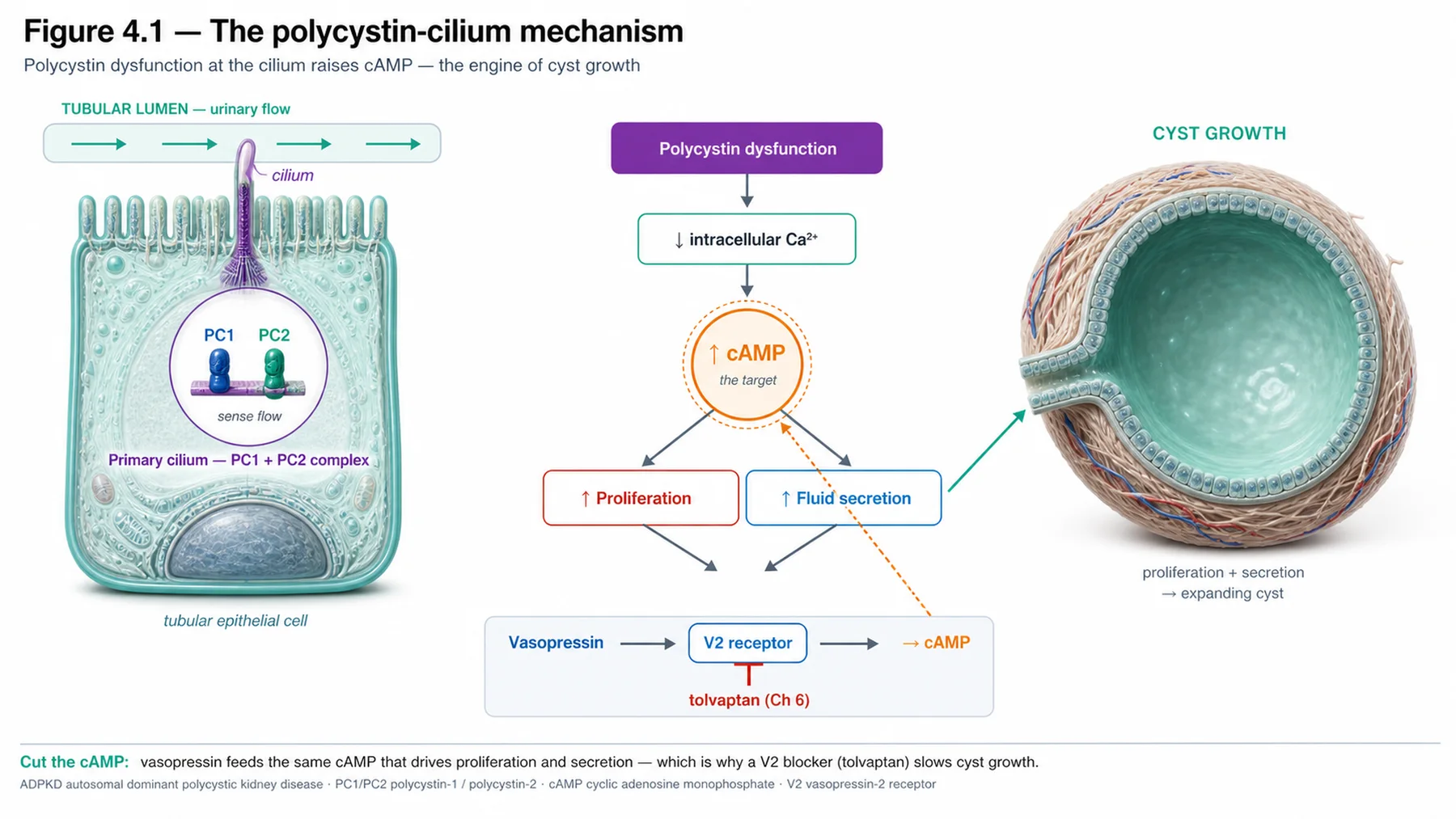
The mechanism of ADPKD is one of the most elegant in nephrology, centred on the primary cilium. The polycystins — polycystin-1 (a large membrane receptor) and polycystin-2 (a calcium-permeable channel of the TRP family) — form a complex that localises to the primary cilium, a tiny antenna-like organelle projecting from the tubular epithelial cell into the lumen. There, the complex senses tubular flow and regulates the cell's behaviour, principally by controlling intracellular calcium (through the polycystin-2 channel). When the polycystins are dysfunctional, this signalling fails: intracellular calcium falls, and — critically — cyclic AMP (cAMP) rises. The raised cAMP has two pro-cystic effects: it drives epithelial cell proliferation (more cells) and transepithelial fluid secretion (cAMP-stimulated chloride and fluid secretion into the lumen). Together, proliferation and fluid secretion are the engine of cyst formation — a tubular segment lined by proliferating, fluid-secreting cells balloons into a fluid-filled cyst. So the chain is: polycystin dysfunction at the cilium → low calcium, high cAMP → proliferation and secretion → cysts. This pathway is not just explanatory; the cAMP node, driven by vasopressin, is the therapeutic target.
— The two-hit model: why cysts are focal
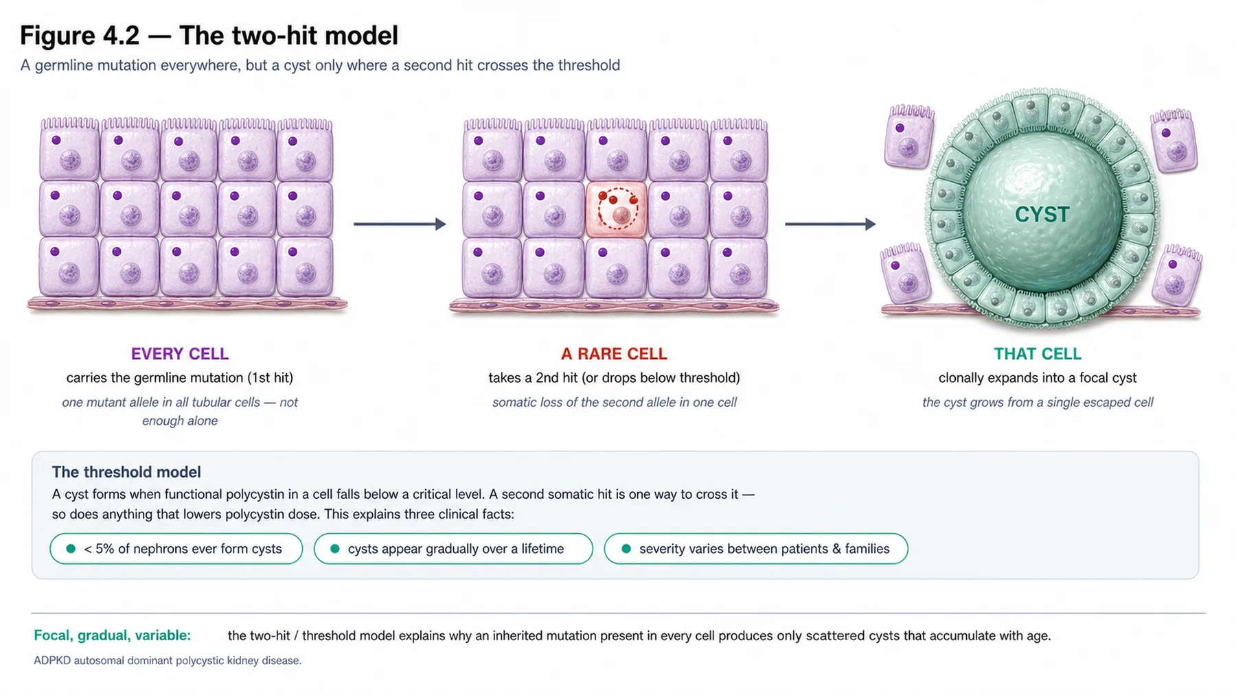
A puzzle the mechanism must explain is why, if the germline mutation is present in every cell of the body, only a small fraction of nephrons (under 5%) actually form cysts — the kidney is not uniformly cystic from birth, and cysts arise focally over time. The answer is the two-hit (or threshold) model. Every cell carries one mutated and one normal allele (the germline heterozygous state), which is insufficient by itself to cause a cyst; cystogenesis is triggered in an individual tubular cell when it sustains a 'second hit' — a somatic mutation inactivating the remaining normal allele — or, more generally, when the functional polycystin level in that cell falls below a critical threshold. Because second hits occur randomly and rarely in individual cells over a lifetime, cysts arise focally and progressively, from scattered nephrons, rather than everywhere at once. This model explains the focal, time-dependent nature of cyst formation, the variability between patients (and even between the two kidneys of one patient), and why the disease, though present genetically from conception, manifests as progressive cyst accumulation over decades. The threshold concept also links to the genotype-phenotype relationship: a more severe (truncating) mutation leaves less functional polycystin, so the threshold is crossed more easily and more often, producing faster disease.
— Cyst growth, vasopressin, and the treatment rationale
Once formed, cysts do not stay static — they grow, and their growth destroys the kidney. A cyst arising from a tubular segment enlarges as its lining cells proliferate and secrete fluid (the cAMP-driven engine), eventually detaching from the parent nephron and becoming an isolated, fluid-filled sac that continues to expand. As cysts grow and multiply over decades, they progressively compress and destroy the surrounding normal parenchyma, provoke interstitial inflammation and fibrosis, and cause the kidneys to enlarge enormously (the rising total kidney volume of the imaging chapter). The key driver to highlight is vasopressin: vasopressin, acting on the V2 receptor in the collecting-duct and cyst epithelium, raises intracellular cAMP — the very signal that drives cyst-cell proliferation and fluid secretion. This makes vasopressin a central promoter of cyst growth, and provides the direct rationale for the disease-modifying treatment developed two chapters on: a vasopressin V2-receptor antagonist (tolvaptan) lowers cyst cAMP and slows cyst growth. So the pathogenesis is not merely academic — the cAMP/vasopressin axis is precisely where the first effective treatment for ADPKD acts.
— The renal natural history
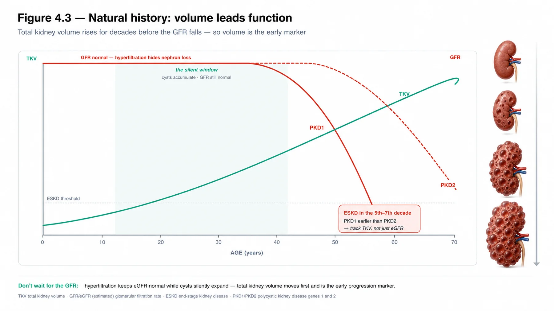
The natural history of ADPKD has a characteristic and clinically important shape. Cysts begin forming from childhood or early adulthood, and the kidneys enlarge progressively over the subsequent decades as cysts accumulate and grow — so the total kidney volume rises steadily and relatively early. The GFR, however, is preserved for a long time: the intact nephrons compensate by hyperfiltration, masking the ongoing destruction, so the GFR remains normal or near-normal for decades even as the kidneys enlarge and parenchyma is lost — and only later, when the compensatory reserve is exhausted, does the GFR begin to decline, often fairly rapidly. This has a crucial consequence: the GFR is a late marker of progression in ADPKD, lagging behind the disease, whereas the total kidney volume rises early and tracks the cyst burden — which is exactly why total kidney volume (not GFR) is the prognostic biomarker that identifies rapid progressors before their GFR falls (the imaging and risk chapters). End-stage kidney disease typically arrives in the fifth to seventh decade, earlier in PKD1 than PKD2. Hypertension appears early and is common (and itself accelerates progression). The lesson is that ADPKD progresses silently — kidneys enlarging, parenchyma lost — long before the GFR reveals it, so progression must be tracked by volume, not function alone.
— The extrarenal manifestations
ADPKD is a systemic disease, and its extrarenal manifestations matter for diagnosis, surveillance, and complications. Polycystic liver disease is the commonest extrarenal feature — cysts in the liver, usually asymptomatic but occasionally causing massive hepatomegaly (more severe in women, influenced by oestrogen). Intracranial (berry) aneurysms are the most feared: they occur more often than in the general population and can rupture catastrophically, so screening (by MR angiography) is offered to patients with a family history of aneurysm or subarachnoid haemorrhage (and in selected high-risk situations). Cardiac valve abnormalities (notably mitral valve prolapse) are more common. And there are connective-tissue-related features — colonic diverticulosis and abdominal wall and inguinal hernias. Recognising these extrarenal features supports the diagnosis (the liver cysts in particular), prompts appropriate screening (the aneurysm question), and anticipates complications. With the genetics, mechanism, and natural history of ADPKD established — the polycystin-cilium defect raising cAMP to drive cyst growth, the two-hit model explaining focal cysts, vasopressin as the treatable driver, and the volume-leads-GFR natural history — the next chapters turn to diagnosing ADPKD, predicting its course by total kidney volume and genetics, and treating it with tolvaptan.
| 04 | PHASE A · LEVEL 4 · ORIENTATION & KNOWLEDGE Reference Tables |
Table 4.1 — Epidemiology and genetics
| Aspect | Detail |
| Frequency | ~1 in 400–1,000 — commonest inherited kidney disease; commonest monogenic ESKD |
| Inheritance | Autosomal dominant — 50% of offspring, both sexes, variable expressivity |
| PKD1 (chr 16, polycystin-1) | ~78% — more severe; ESKD ~mid-to-late 50s; truncating worst |
| PKD2 (chr 4, polycystin-2) / others | ~15% — milder, ESKD ~70s; rarer genes (GANAB, DNAJB11), some undetected |
Table 4.2 — The polycystin-cilium mechanism
| Element | Detail |
| Polycystin-1 | Large membrane receptor at the primary cilium (mechano/flow sensing) |
| Polycystin-2 | Calcium-permeable TRP channel — partners PC1 |
| Dysfunction | ↓ intracellular calcium, ↑ cAMP |
| Consequence | cAMP drives epithelial proliferation + fluid secretion → cysts |
Table 4.3 — The two-hit (threshold) model
| Point | Detail |
| Germline state | Heterozygous mutation in every cell — insufficient alone |
| Second hit / threshold | Somatic inactivation of the normal allele, or functional polycystin below threshold |
| Result | Cystogenesis triggered focally in individual cells (< 5% of nephrons) |
| Explains | Focal, time-dependent cysts; inter-patient and inter-kidney variability |
Table 4.4 — Cyst growth and vasopressin
| Aspect | Detail |
| Cyst growth | Proliferation + cAMP-driven fluid secretion → cysts enlarge, detach |
| Parenchymal damage | Compression, inflammation, fibrosis → kidney enlargement, function loss |
| Vasopressin | V2 receptor → ↑ cAMP in cyst epithelium → promotes cyst growth |
| Treatment rationale | V2-receptor antagonist (tolvaptan) → ↓ cAMP → slows cyst growth (Chapter 6) |
Table 4.5 — Renal natural history
| Phase | Detail |
| Cyst formation | From childhood/early adulthood; kidneys enlarge progressively (TKV rises early) |
| GFR preserved | Compensatory hyperfiltration masks loss for decades — GFR is a LATE marker |
| Decline | GFR falls once reserve exhausted; ESKD typically 5th–7th decade (PKD1 earlier) |
| Hypertension | Early and common — accelerates progression |
Table 4.6 — Extrarenal manifestations
| Manifestation | Detail |
| Polycystic liver disease | Commonest extrarenal; usually asymptomatic; more severe in women |
| Intracranial aneurysms | Increased risk; screen if family history of aneurysm/SAH |
| Cardiac valves | Mitral valve prolapse and others |
| Other | Colonic diverticulosis; abdominal wall and inguinal hernias |
| 05 | PHASE B · LEVEL 5 · VISUALISE & MAP Imaging & Flowchart Specifications |
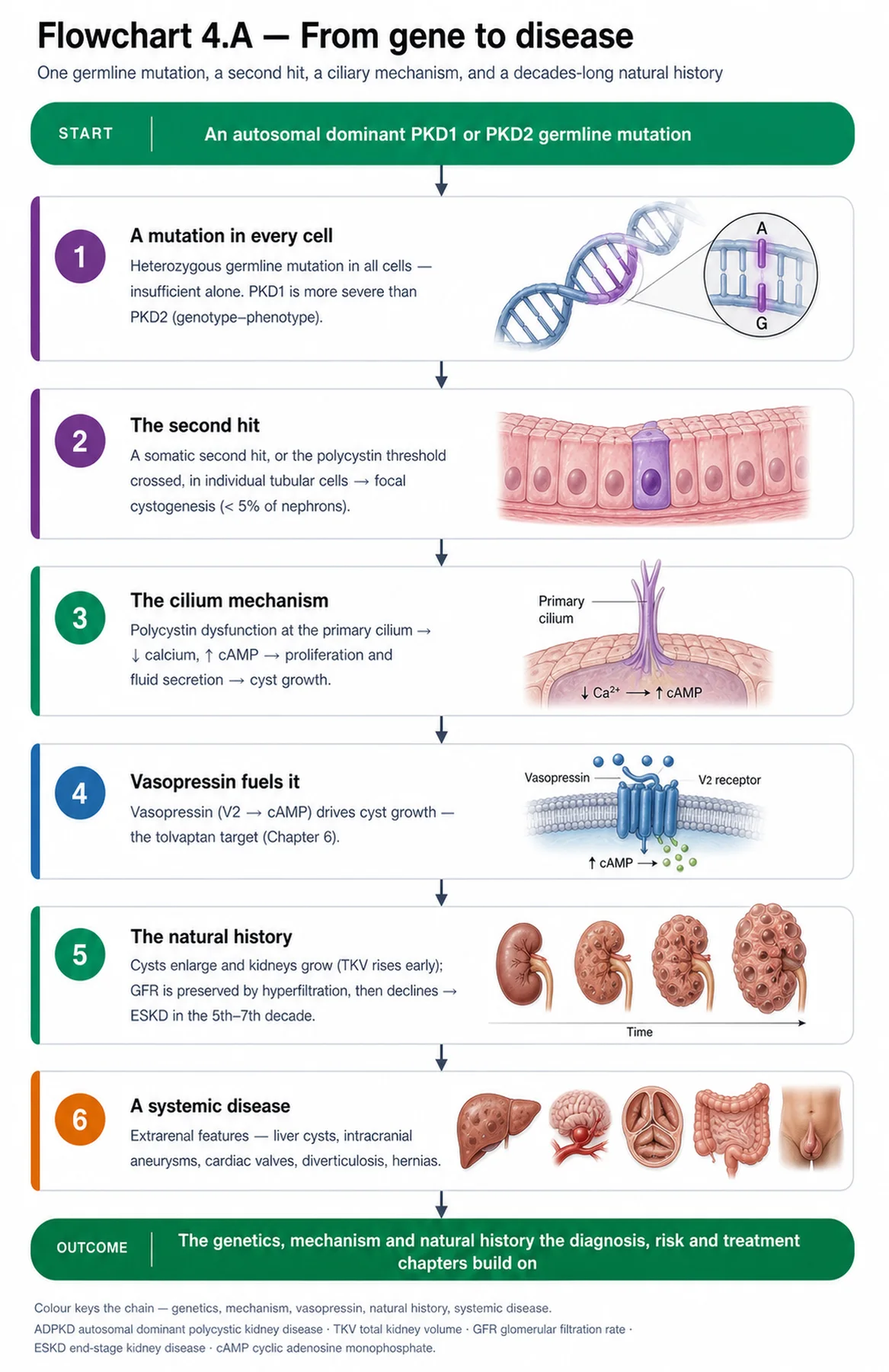
| 06 | PHASE B · LEVEL 6 · VISUALISE & MAP Concept Maps |
Each chain runs from mechanism to a named clinical insight; read the arrows as “leads to.”
Genetics / genotype-phenotype. PKD1 (78%, polycystin-1) or PKD2 (15%, polycystin-2) → PKD1 more severe (ESKD ~2 decades earlier; truncating worst) → ACTION: identify the gene/mutation to help predict the course.
Polycystin-cilium mechanism. Polycystin dysfunction at the primary cilium → ↓ intracellular calcium, ↑ cAMP → epithelial proliferation + fluid secretion → cysts → ACTION: recognise the cAMP node as the therapeutic target.
Two-hit model. Germline heterozygous mutation everywhere + a second somatic hit (or threshold crossed) in individual cells → focal cysts (< 5% of nephrons) → ACTION: explain the focal, progressive, variable nature of cysts.
Cyst growth / vasopressin. cAMP-driven proliferation + fluid secretion → cyst growth and parenchymal destruction; vasopressin (V2 → cAMP) promotes it → ACTION: target vasopressin (tolvaptan) to slow cyst growth.
Natural history. Cysts accumulate → TKV rises early, but compensatory hyperfiltration preserves GFR for decades → GFR is a late marker → ACTION: track progression by total kidney volume, not GFR alone.
| 07 | PHASE B · LEVEL 7 · VISUALISE & MAP Decision Pathways |
| R1 | IF counselling an ADPKD patient, THEN recognise the autosomal dominant inheritance — 50% of offspring at risk, both sexes, vertical transmission. |
| R2 | IF the gene is known, THEN use it to predict the course — PKD1 (especially truncating) more severe than PKD2. |
| R3 | IF explaining cyst formation, THEN attribute it to polycystin dysfunction at the cilium raising cAMP and driving proliferation and fluid secretion. |
| R4 | IF asked why only some nephrons form cysts, THEN invoke the two-hit/threshold model — focal cystogenesis on a germline background. |
| R5 | IF considering disease-modifying treatment, THEN recall that vasopressin (V2 → cAMP) drives cyst growth — the rationale for tolvaptan. |
| R6 | IF monitoring progression, THEN track total kidney volume, which rises early, rather than relying on GFR, which is preserved for decades. |
| R7 | IF a patient has a family history of intracranial aneurysm or subarachnoid haemorrhage, THEN offer aneurysm screening. |
| R8 | IF an ADPKD patient is hypertensive, THEN treat it — it appears early and accelerates progression. |
| 08 | PHASE C · LEVEL 8 · CLINICAL REASONING Clinical Cases |
| CASE 1 | WHICH GENE, WHICH COURSE Genotype predicts severity PKD1 versus PKD2 |
Presentation
Two ADPKD patients ask about their prognosis. One has a PKD1 truncating mutation; the other has PKD2. A trainee gives them the same generic outlook.
❖ Pause and reflect Should these two patients expect the same course? |
Analysis
No — the gene (and mutation type) predicts a substantially different course. ADPKD has a clear genotype-phenotype relationship: PKD1 disease is more severe than PKD2, reaching end-stage kidney disease roughly two decades earlier (median in the mid-to-late 50s for PKD1 versus the 70s for PKD2), and within PKD1, truncating mutations are the most severe. So the patient with a PKD1 truncating mutation has a more aggressive course and earlier expected kidney failure than the patient with PKD2. Giving them the same generic outlook ignores this prognostic information. The gene therefore matters not only for diagnosis but for prognosis and for decisions such as the intensity of monitoring and the consideration of disease-modifying treatment.
Plan
Use the genotype to individualise the prognosis — a more aggressive course for the PKD1 truncating mutation, a milder one for PKD2 — and tailor monitoring and treatment consideration accordingly. Let the gene inform the prognosis.
Teaching point
ADPKD has a genotype-phenotype relationship — PKD1 (especially truncating) is more severe than PKD2, reaching ESKD ~two decades earlier; the gene informs prognosis.
Cross-reference
Exercises rules R1 and R2; the genetics concept map; Table 4.1; risk prediction in Chapter 5.
| CASE 2 | WHY VASOPRESSIN MATTERS The cAMP driver The treatment rationale |
Presentation
A patient with ADPKD asks how the disease-modifying treatment they have heard about could possibly work, given that the disease is genetic. A trainee is unsure how to explain the rationale.
❖ Pause and reflect How can a drug slow a genetic cystic disease? |
Analysis
By targeting the downstream driver of cyst growth, even though the underlying defect is genetic. The polycystin dysfunction raises intracellular cAMP, and cAMP drives the epithelial proliferation and fluid secretion that grow the cysts; vasopressin, acting on the V2 receptor in the cyst epithelium, is a major stimulus of that cAMP. So although the genetic defect cannot be corrected, the cAMP/vasopressin axis through which it grows cysts can be interrupted: a vasopressin V2-receptor antagonist (tolvaptan) lowers cyst cAMP and slows cyst growth and the decline in function. This is the elegant link between mechanism and treatment — the drug works not by fixing the gene but by blocking the vasopressin-cAMP signal that the genetic defect exploits to grow cysts. The mechanism explains the therapy.
Plan
Explain that the genetic defect grows cysts through a cAMP signal stimulated by vasopressin, and that blocking vasopressin's V2 receptor (tolvaptan) lowers cyst cAMP and slows growth — a downstream, not genetic, treatment (developed in Chapter 6). The mechanism is the rationale.
Teaching point
Vasopressin (V2 → cAMP) drives cyst growth — so a vasopressin antagonist (tolvaptan) slows ADPKD by interrupting the downstream driver, not by fixing the gene.
Cross-reference
Exercises rule R5; the polycystin and vasopressin concept maps; Figure 4.1; Tables 4.2, 4.4; tolvaptan in Chapter 6.
| CASE 3 | VOLUME BEFORE FUNCTION GFR is a late marker Natural history |
Presentation
A young ADPKD patient with markedly enlarged, cystic kidneys but a normal eGFR is reassured that 'the kidneys are working fine' and that monitoring can be infrequent.
❖ Pause and reflect Does a normal eGFR mean the disease is not progressing? |
Analysis
No — a normal eGFR in ADPKD is reassuring only superficially, because the GFR is a late marker that lags behind the disease. The intact nephrons compensate by hyperfiltration, maintaining a normal or near-normal GFR for decades even as cysts accumulate, the kidneys enlarge, and parenchyma is progressively destroyed — so the markedly enlarged cystic kidneys here signal active, ongoing progression despite the normal eGFR. The total kidney volume rises early and tracks the cyst burden, whereas the GFR falls only once the compensatory reserve is exhausted. Reassuring this patient on the eGFR alone, and reducing monitoring, misreads the natural history: progression should be tracked by total kidney volume (the prognostic biomarker), and a young patient with already markedly enlarged kidneys may well be a rapid progressor needing closer follow-up and treatment consideration.
Plan
Do not be reassured by the normal eGFR; recognise the enlarged cystic kidneys as evidence of progression, track total kidney volume as the early marker, and assess this potentially rapid progressor for closer monitoring and treatment. Track volume, not GFR alone, in ADPKD.
Teaching point
In ADPKD, GFR is preserved for decades by hyperfiltration — a late marker; total kidney volume rises early, so track volume, not GFR alone.
Cross-reference
Exercises rule R6; the natural-history concept map; Figure 4.3; Table 4.5; TKV in Chapter 3 and risk in Chapter 5.
| CASE 4 | BEYOND THE KIDNEY The systemic disease Extrarenal manifestations |
Presentation
An ADPKD patient with a family history of a relative who died of a 'brain bleed' is managed for the kidneys alone, with no consideration of the extrarenal disease.
❖ Pause and reflect What extrarenal issue must be addressed here? |
Analysis
Intracranial aneurysm screening, given the family history. ADPKD is a systemic disease, and intracranial (berry) aneurysms are its most feared extrarenal manifestation — more common than in the general population, with the risk of catastrophic rupture (subarachnoid haemorrhage). The family history of a relative who died of a 'brain bleed' (likely a ruptured aneurysm) places this patient in the group for whom aneurysm screening (by MR angiography) is offered, because the risk is familial. Managing the kidneys alone misses this. More broadly, the systemic features — polycystic liver disease (the commonest extrarenal feature), cardiac valve abnormalities, diverticulosis, and hernias — should be recognised, but the aneurysm screening is the pressing, potentially life-saving action here.
Plan
Offer intracranial aneurysm screening (MR angiography) given the family history of probable aneurysmal haemorrhage, and recognise the broader extrarenal disease (liver cysts, valves, diverticulosis, hernias). Address the systemic disease, especially aneurysm screening when familial.
Teaching point
ADPKD is systemic — offer intracranial aneurysm screening when there is a family history of aneurysm or subarachnoid haemorrhage; recognise liver cysts and the other extrarenal features.
Cross-reference
Exercises rule R7; Table 4.6.
| 09 | PHASE C · LEVEL 9 · CLINICAL REASONING Clinical Implications |
One triad per mechanism the narrative exposed: the physiology, why it matters, and the clinical move.
MECHANISM ADPKD is caused by PKD1 or PKD2 mutations, with PKD1 more severe. |
WHY IT MATTERS The gene and mutation type predict the rate of progression. |
ACTION Identify the gene to individualise the prognosis. |
MECHANISM Polycystin dysfunction at the cilium lowers calcium and raises cAMP. |
WHY IT MATTERS cAMP drives the proliferation and fluid secretion that form cysts. |
ACTION Recognise the cAMP node as the therapeutic target. |
MECHANISM Cystogenesis requires a second hit on a germline-mutated background. |
WHY IT MATTERS This explains the focal, progressive, and variable nature of cysts. |
ACTION Use the two-hit model to explain the disease's course and variability. |
MECHANISM Vasopressin raises cyst-cell cAMP through the V2 receptor. |
WHY IT MATTERS It is a major driver of cyst growth. |
ACTION Target vasopressin with a V2-receptor antagonist to slow cyst growth. |
MECHANISM Compensatory hyperfiltration preserves the GFR for decades while cysts grow. |
WHY IT MATTERS The GFR is a late marker, lagging behind the disease. |
ACTION Track progression by total kidney volume, not GFR alone. |
| 10 | PHASE C · LEVEL 10 · CLINICAL REASONING Clinical Pearls |
| ADPKD: commonest inherited kidney disease; commonest monogenic cause of ESKD. | Autosomal dominant — 50% of offspring, both sexes, variable expressivity. |
| PKD1 (chr 16, polycystin-1) ~78%; PKD2 (chr 4, polycystin-2) ~15%. | PKD1 more severe than PKD2 (ESKD ~2 decades earlier); truncating worst. |
| Polycystins localise to the primary cilium (PC1 receptor, PC2 calcium channel). | Dysfunction → ↓ intracellular calcium, ↑ cAMP. |
| cAMP drives epithelial proliferation + fluid secretion → cysts. | Two-hit/threshold model → focal cysts (< 5% of nephrons) despite germline mutation. |
| Cysts enlarge, detach, secrete fluid → destroy parenchyma → kidney enlargement. | Vasopressin (V2 → cAMP) drives cyst growth — the tolvaptan rationale. |
| Cysts form early; kidneys enlarge (TKV rises early). | GFR preserved for decades by hyperfiltration — a LATE marker. |
| Track progression by total kidney volume, not GFR alone. | ESKD typically 5th–7th decade (PKD1 earlier); hypertension early and common. |
| Extrarenal: liver cysts (commonest), intracranial aneurysms, valves, diverticulosis, hernias. | Screen for aneurysms if family history of aneurysm/SAH. |
| 11 | PHASE D · LEVEL 11 · SAFETY & EVIDENCE Red Flags & Never-Do |
Panel A — Red flags
| ▲ | A normal eGFR in ADPKD taken as evidence of no progression — GFR is a late marker; track total kidney volume. |
| ▲ | A PKD1 truncating mutation — the most aggressive course; expect earlier ESKD. |
| ▲ | A family history of aneurysm or subarachnoid haemorrhage — offer intracranial aneurysm screening. |
| ▲ | Early hypertension in ADPKD — common and progression-accelerating; treat it. |
| ▲ | Managing the kidneys alone — ADPKD is systemic (liver, aneurysms, valves). |
Panel B — Never do
| ✖ NEVER — reassure an ADPKD patient on a normal eGFR alone — it is a late marker. |
| ✖ NEVER — give every ADPKD patient the same prognosis regardless of the gene. |
| ✖ NEVER — overlook aneurysm screening when there is a familial history of haemorrhage. |
| ✖ NEVER — treat ADPKD as a purely renal disease. |
| 12 | PHASE D · LEVEL 12 · SAFETY & EVIDENCE Common Pitfalls |
Pitfall 1 — Reassuring on eGFR
| ✖ | WRONG Reassuring an ADPKD patient because the eGFR is normal. |
| ✓ | RIGHT Tracking total kidney volume as the early marker. |
| ✉ | WHY Compensatory hyperfiltration preserves GFR for decades. |
Pitfall 2 — One prognosis for all
| ✖ | WRONG Giving every ADPKD patient the same generic outlook. |
| ✓ | RIGHT Using the gene/mutation to individualise the prognosis. |
| ✉ | WHY PKD1 (especially truncating) is more severe than PKD2. |
Pitfall 3 — 'A genetic disease can't be treated'
| ✖ | WRONG Assuming nothing can slow a genetic cystic disease. |
| ✓ | RIGHT Recognising the cAMP/vasopressin driver as a treatable downstream target. |
| ✉ | WHY Tolvaptan blocks the vasopressin-cAMP signal that grows cysts. |
Pitfall 4 — Missing aneurysm screening
| ✖ | WRONG Ignoring a family history of subarachnoid haemorrhage. |
| ✓ | RIGHT Offering intracranial aneurysm screening. |
| ✉ | WHY Aneurysms are more common in ADPKD and cluster in families. |
Pitfall 5 — Renal tunnel vision
| ✖ | WRONG Managing ADPKD as a purely renal disease. |
| ✓ | RIGHT Recognising and addressing the systemic manifestations. |
| ✉ | WHY ADPKD affects liver, brain vessels, heart valves, and connective tissue. |
| 13 | PHASE D · LEVEL 13 · SAFETY & EVIDENCE Evidence Grading |
GRADE A HIGH CONFIDENCE The effect is real and the estimate is stable. RCTs at low risk of bias; multiple concordant prospective cohorts; meta-analyses. |
GRADE B MODERATE CONFIDENCE The effect is likely real but may shift with new data. Observational studies, registries, mechanistic human studies. |
GRADE C LOW CONFIDENCE Rests on physiology, reasoning, or consensus rather than outcomes. Pathophysiological reasoning; extrapolation; consensus without outcomes. |
Graded statements (by evidence type)
| Statement | Grade | Basis (evidence type) |
| ADPKD is the commonest inherited kidney disease and monogenic cause of ESKD. | A | Epidemiological data |
| PKD1 disease is more severe than PKD2. | A | Cohort and genotype-phenotype data |
| Polycystin dysfunction raises cAMP, driving cyst growth. | A | Mechanistic studies |
| A two-hit/threshold mechanism explains focal cystogenesis. | A | Molecular studies |
| Vasopressin promotes cyst growth via V2-receptor cAMP signalling. | A | Mechanistic and clinical data |
| GFR is preserved for decades by compensatory hyperfiltration. | A | Natural-history studies |
| Intracranial aneurysms are more common in ADPKD. | A | Cohort data |
| 18 | PHASE F · LEVEL 18 · APPLY & TEST Cheat Sheet |
| ADPKD: commonest inherited kidney disease + monogenic ESKD cause. | Autosomal dominant; 50% of offspring; variable expressivity. |
| PKD1 (polycystin-1) ~78%; PKD2 (polycystin-2) ~15%. | PKD1 > PKD2 in severity (ESKD ~2 decades earlier); truncating worst. |
| Polycystins at the primary cilium (PC1 receptor, PC2 Ca channel). | Dysfunction → ↓ calcium, ↑ cAMP. |
| cAMP → proliferation + fluid secretion → cysts. | Two-hit/threshold → focal cysts (< 5% of nephrons). |
| Cysts grow/detach/secrete → destroy parenchyma. | Vasopressin (V2 → cAMP) drives growth — tolvaptan target. |
| Cysts early; TKV rises early. | GFR preserved by hyperfiltration — LATE marker. |
| Track progression by TKV, not GFR alone. | ESKD 5th–7th decade (PKD1 earlier); early hypertension. |
| Extrarenal: liver cysts (commonest), aneurysms, valves, diverticulosis, hernias. | Aneurysm screening if family history of aneurysm/SAH. |
| 19 | PHASE F · LEVEL 19 · APPLY & TEST Flashcards |
| CARD 1 | Q. What is the epidemiology and inheritance of ADPKD? A. It affects roughly one in 400 to 1,000 people — the commonest inherited kidney disease and the commonest monogenic cause of end-stage kidney disease — and is autosomal dominant, so each child of an affected parent has a 50% chance of inheriting it, with variable expressivity. DETAILED. Both sexes are affected, with vertical transmission. CLINICAL. Counsel on the 50% offspring risk. |
| CARD 2 | Q. What genes cause ADPKD, and how do they differ? A. PKD1 (chromosome 16, polycystin-1) causes about 78% and PKD2 (chromosome 4, polycystin-2) about 15%, with rarer genes for the rest; PKD1 disease is more severe than PKD2, reaching end-stage disease roughly two decades earlier, and PKD1 truncating mutations are the most severe. DETAILED. The gene predicts the course. CLINICAL. Use the genotype to individualise the prognosis. |
| CARD 3 | Q. How does polycystin dysfunction cause cysts? A. The polycystins (polycystin-1 a receptor, polycystin-2 a calcium channel) localise to the primary cilium and sense tubular flow; their dysfunction lowers intracellular calcium and raises cAMP, which drives epithelial proliferation and fluid secretion — the engine of cyst formation and growth. DETAILED. The cAMP node is the therapeutic target. CLINICAL. Recognise the calcium-cAMP pathway as the cyst driver. |
| CARD 4 | Q. What is the two-hit model, and what does it explain? A. Every cell carries the germline heterozygous mutation (insufficient alone); cystogenesis is triggered in an individual cell by a second somatic hit, or by functional polycystin falling below a critical threshold — explaining why fewer than 5% of nephrons form cysts and why cysts arise focally, progressively, and variably. DETAILED. It reconciles a universal mutation with focal cysts. CLINICAL. Use it to explain the focal, progressive, variable disease. |
| CARD 5 | Q. How does vasopressin drive cyst growth, and why does it matter? A. Vasopressin acts on the V2 receptor in the cyst epithelium to raise intracellular cAMP — the signal that drives cyst-cell proliferation and fluid secretion — so it is a major promoter of cyst growth, providing the rationale for the V2-receptor antagonist tolvaptan. DETAILED. The genetic defect grows cysts through this treatable axis. CLINICAL. Target vasopressin to slow cyst growth (Chapter 6). |
| CARD 6 | Q. Why is the GFR preserved for decades in ADPKD? A. Because the intact nephrons compensate by hyperfiltration, masking the ongoing destruction, so the GFR stays normal or near-normal for decades even as the kidneys enlarge and parenchyma is lost — making the GFR a late marker that lags behind the disease. DETAILED. Total kidney volume rises early and tracks the cyst burden. CLINICAL. Track progression by total kidney volume, not GFR alone. |
| CARD 7 | Q. What is the renal natural history of ADPKD? A. Cysts form from childhood or early adulthood, the kidneys enlarge progressively (rising total kidney volume), the GFR is preserved for decades then declines once compensatory reserve is exhausted, and end-stage disease typically occurs in the fifth to seventh decade (earlier in PKD1); hypertension appears early and accelerates progression. DETAILED. Volume leads function. CLINICAL. Expect silent progression — monitor by volume and treat the hypertension. |
| CARD 8 | Q. What are the major extrarenal manifestations of ADPKD? A. Polycystic liver disease (the commonest, more severe in women), intracranial (berry) aneurysms (with the risk of subarachnoid haemorrhage), cardiac valve abnormalities (mitral valve prolapse), colonic diverticulosis, and abdominal wall and inguinal hernias. DETAILED. ADPKD is a systemic disease. CLINICAL. Recognise the extrarenal features and screen for aneurysms when there is a familial history. |
| 20 | PHASE F · LEVEL 20 · APPLY & TEST One-Minute Preceptor |
| SCENE 1 | The intern reassuring on eGFR |
GET A COMMITMENT. “You've reassured this young ADPKD patient with hugely enlarged kidneys because the eGFR is normal — is that right?”
PROBE FOR EVIDENCE. “The eGFR is fine” — ask: “What keeps the GFR normal for decades in ADPKD, and what is rising in the meantime?”
TEACH A GENERAL RULE. Compensatory hyperfiltration preserves the GFR for decades, so it is a late marker; total kidney volume rises early and tracks the disease — monitor by volume, not GFR.
REINFORCE WHAT WAS RIGHT. Checking the eGFR was reasonable.
CORRECT A MISTAKE. Track total kidney volume; the enlarged kidneys signal active progression.
| SCENE 2 | The resident doubting treatment |
GET A COMMITMENT. “You've told this patient nothing can be done because ADPKD is genetic — is that accurate?”
PROBE FOR EVIDENCE. “The gene can't be fixed” — ask: “What downstream signal grows the cysts, and what stimulates it?”
TEACH A GENERAL RULE. The genetic defect grows cysts through cAMP, which vasopressin stimulates — so a vasopressin antagonist (tolvaptan) slows the disease by blocking the driver, even without fixing the gene.
REINFORCE WHAT WAS RIGHT. Recognising the genetic basis was correct.
CORRECT A MISTAKE. Explain the treatable vasopressin-cAMP axis (Chapter 6).
| 22 | PHASE F · LEVEL 22 · APPLY & TEST Board-Style Questions |
| Q 01 | ADPKD is most often caused by mutations in: |
| A | PKHD1 |
| B | PKD1 (polycystin-1) |
| C | COL4A5 |
| D | GLA |
Rationale PKD1 (polycystin-1) causes ~78% of ADPKD (Table 4.1). A is ARPKD; C is Alport; D is Fabry. |
| Q 02 | Compared with PKD2, PKD1 disease: |
| A | Is milder |
| B | Is more severe, reaching ESKD roughly two decades earlier |
| C | Is identical |
| D | Never reaches ESKD |
Rationale PKD1 is more severe with earlier ESKD; truncating mutations worst (case 1, Table 4.1). A, C, and D are wrong. |
| Q 03 | Polycystin dysfunction promotes cyst formation by: |
| A | Raising intracellular calcium |
| B | Lowering calcium and raising cAMP, driving proliferation and fluid secretion |
| C | Blocking fluid secretion |
| D | Stopping proliferation |
Rationale Dysfunction lowers calcium and raises cAMP, the engine of cyst growth (Figure 4.1, Table 4.2). A, C, and D are the opposite. |
| Q 04 | Why do only a small fraction of nephrons form cysts despite a germline mutation in every cell? |
| A | The mutation is not really present |
| B | A second somatic hit (or threshold crossing) is needed in individual cells |
| C | Cysts are congenital and fixed |
| D | The kidney repairs them |
Rationale The two-hit/threshold model explains focal cystogenesis (Figure 4.2, Table 4.3). A, C, and D are incorrect. |
| Q 05 | Vasopressin promotes cyst growth in ADPKD by: |
| A | Lowering cAMP |
| B | Raising cyst-cell cAMP via the V2 receptor |
| C | Blocking the polycystins |
| D | Reducing fluid secretion |
Rationale Vasopressin (V2 → cAMP) drives cyst growth — the tolvaptan rationale (case 2, Table 4.4). A, C, and D are wrong. |
| Q 06 | Why is the GFR preserved for decades in ADPKD? |
| A | The cysts improve function |
| B | Compensatory hyperfiltration of intact nephrons masks the loss |
| C | There is no nephron loss |
| D | The GFR is never normal |
Rationale Hyperfiltration preserves the GFR, making it a late marker (case 3, Figure 4.3, Table 4.5). A, C, and D are incorrect. |
| Q 07 | Progression in ADPKD is best tracked by: |
| A | GFR alone |
| B | Total kidney volume (it rises early) |
| C | Blood pressure alone |
| D | Urine output |
Rationale TKV rises early while the GFR is preserved (Figure 4.3, rule R6). A, C, and D are inadequate. |
| Q 08 | Which is the commonest extrarenal manifestation of ADPKD? |
| A | Intracranial aneurysm |
| B | Polycystic liver disease |
| C | Diverticulosis |
| D | Mitral valve prolapse |
Rationale Polycystic liver disease is the commonest extrarenal feature (case 4, Table 4.6). A, C, and D occur but are less common. |