Chapter Preamble
Signals declared
Sig-D — Diagnostic (primary). Use imaging, biopsy, and biomarkers — alongside genetics — to diagnose and characterise inherited and cystic kidney disease.
Sig-V — Evidence-dense (strong). The validated ultrasound criteria, total kidney volume as a prognostic biomarker, and the changing role of biopsy in the genetic era — graded and reflected on.
Levels populated and omitted
Populated (17): L1–L5, L7, L8, L10–L14, L18–L22. The evidence signal fires the absolute-risk table (L14) and the reflective prompts (L21); the diagnostic signal drives the tables, rules, cases, pitfalls, and board items.
L6 / L9 mechanism levels — omitted. No Sig-M; this is a diagnostic-tools and evidence chapter.
L15 / L16 preference-sensitive map and SDM scripts — omitted. No Sig-E; selecting and interpreting the diagnostic tools is a technical and evidential matter.
L17 documentation templates — omitted. No Sig-P/T; the management templates belong with the disease-specific chapters.
| 01 | PHASE A · LEVEL 1 · ORIENTATION & KNOWLEDGE Learning Objectives |
By the end of this chapter you should be able to:
Describe the imaging modalities and their roles in inherited and cystic kidney disease.
Apply the ultrasound criteria for ADPKD and recognise their limitations.
Explain total kidney volume as a prognostic biomarker and the role of MRI.
State the changing role of biopsy in the genetic era and when it is still needed.
Recognise the characteristic biopsy findings of Alport and Fabry disease.
List the established and emerging biomarkers.
Explain how imaging, biopsy, biomarkers, and genetics integrate.
Summarise the evidence behind the ultrasound criteria and total kidney volume.
| 02 | PHASE A · LEVEL 2 · ORIENTATION & KNOWLEDGE Executive Summary |
The diagnostic toolkit for inherited and cystic kidney disease comprises imaging, biopsy, biomarkers, and — increasingly central — genetics, used together.
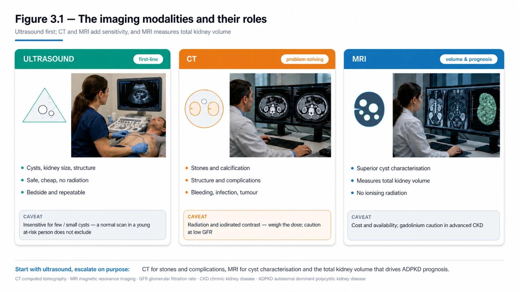
Ultrasound is the first-line imaging modality: safe, cheap, and free of radiation and contrast, detecting cysts, kidney size, and structure, and forming the basis of the validated age-based ultrasound criteria for diagnosing ADPKD in at-risk individuals.
Ultrasound is insensitive for the few small cysts of young at-risk patients, where genetics or MRI may be needed.
CT and MRI are more sensitive for cysts, stones, structure, and complications, with MRI the gold standard for measuring total kidney volume.
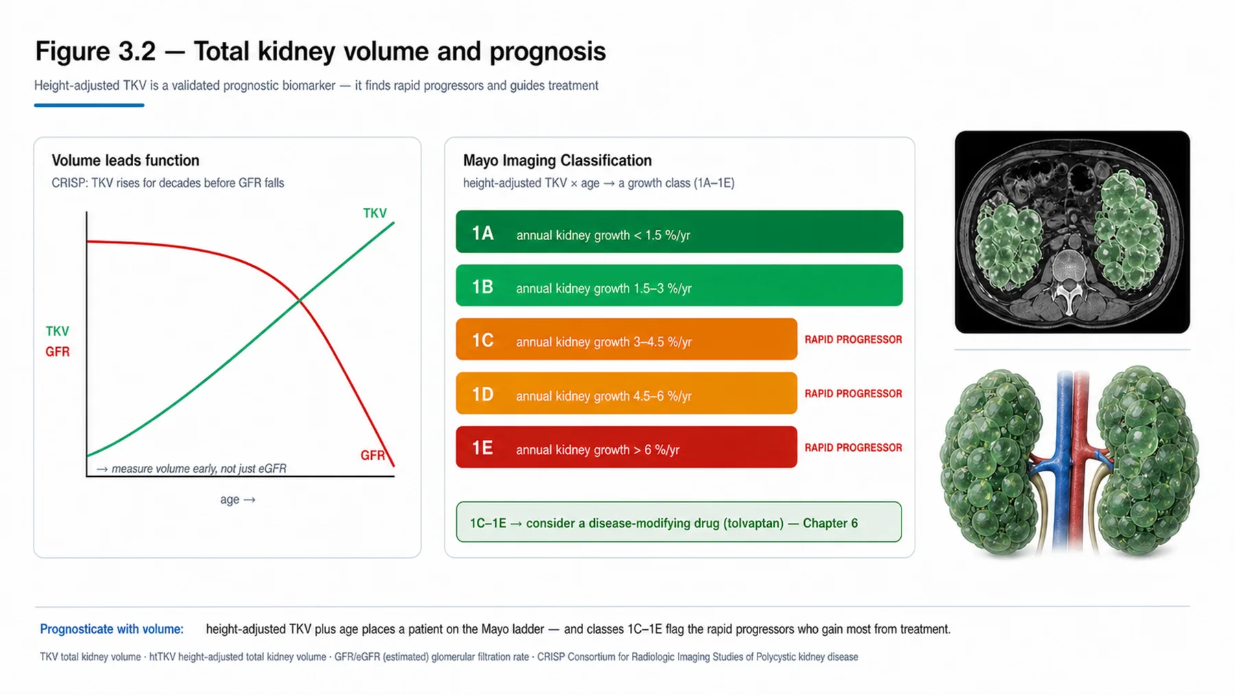
Total kidney volume is the validated prognostic biomarker in ADPKD — height-adjusted and combined with age in the Mayo classification — and is used to identify rapid progressors and to select patients for treatment.
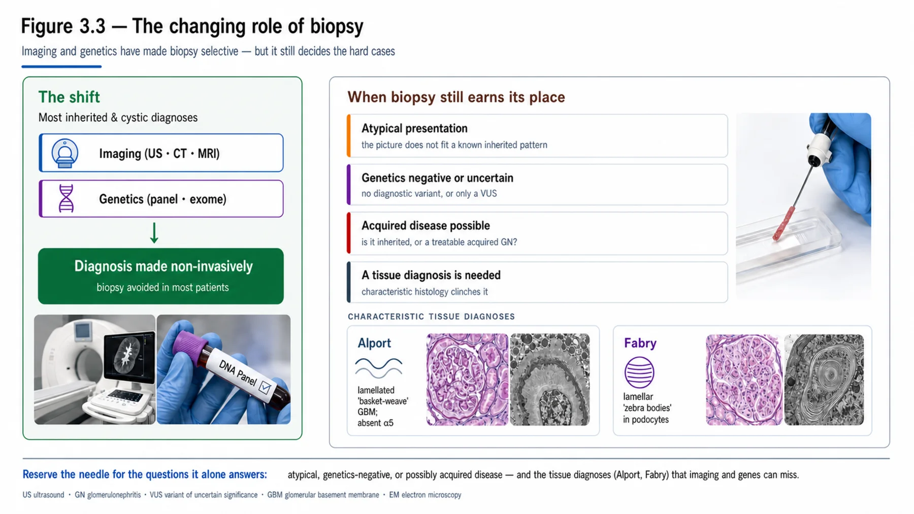
Biopsy is less central in inherited disease than in glomerular disease, because imaging and genetics often make the diagnosis non-invasively.
Biopsy remains valuable in atypical presentations, when genetics is negative or uncertain, when an acquired disease is in the differential, and for specific tissue diagnoses — the lamellated basement membrane and absent collagen IV of Alport, the zebra bodies of Fabry.
Genetic testing has reduced, but not eliminated, the need for biopsy.
Established biomarkers include the eGFR, proteinuria and albuminuria, urinalysis (haematuria in Alport), specific biochemistry (the electrolyte patterns of tubulopathies, urine oxalate, urine cystine), and enzyme assays (alpha-galactosidase A for Fabry).
Emerging biomarkers and, above all, the genetic diagnosis itself — the definitive molecular marker — complete the picture.
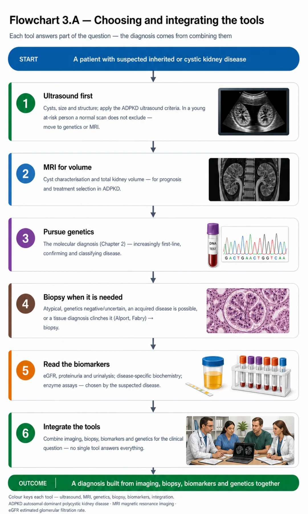
The tools are complementary: imaging for structure and cysts, biopsy for tissue when needed, biomarkers for function and prognosis, and genetics for the molecular diagnosis.
Choosing and integrating them — not relying on any one — is the diagnostic skill.
| 03 | PHASE A · LEVEL 3 · ORIENTATION & KNOWLEDGE Main Narrative |
Diagnosing inherited and cystic kidney disease draws on a toolkit — imaging, biopsy, biomarkers, and genetics — each contributing a different kind of information. The previous chapter covered genetics; this one covers the rest and shows how they fit together. The balance among them has shifted: genetics, once a last resort, is now often central, while biopsy, once routine, is now selective. But each tool retains a role, and the skill is choosing and integrating them rather than relying on any one.
— Imaging: ultrasound first
Ultrasound is the first-line imaging modality for inherited and cystic kidney disease, because it is safe, inexpensive, widely available, and free of radiation and contrast — and it detects what matters most: cysts, kidney size, echogenicity, structure, and nephrocalcinosis. Its central role in this volume is the diagnosis of ADPKD in at-risk individuals, where validated age-based ultrasound criteria (the Pei/Ravine criteria) specify the number of cysts required to make or exclude the diagnosis at different ages in someone with a family history — more cysts being required at older ages, because cysts accumulate over time, and specific absence criteria allowing exclusion in older at-risk relatives (important for living-donor evaluation). The key limitation of ultrasound is its insensitivity for the few, small cysts of a young at-risk individual: a normal ultrasound in a young person does not exclude ADPKD, so genetics or MRI may be needed to diagnose or exclude it (again, important before accepting a young related donor). Ultrasound is the workhorse, but its limits in the young must be respected.
— CT, MRI, and total kidney volume
When ultrasound is insufficient, cross-sectional imaging adds sensitivity and specific capabilities. CT is sensitive for cysts, stones, structure, and complications, but carries radiation and (with iodinated contrast) caution in CKD. MRI avoids radiation and is particularly valuable for characterising cysts (haemorrhage, complexity, features suspicious for malignancy) and — most importantly — for measuring total kidney volume, the key prognostic biomarker in ADPKD. Total kidney volume (TKV) deserves emphasis: the kidneys in ADPKD enlarge as cysts grow, and the TKV — height-adjusted and combined with age — predicts the rate of GFR decline (established by the CRISP study) and is captured in the Mayo imaging classification, which stratifies patients into risk classes. This matters practically because it identifies the rapid progressors who stand to benefit most from disease-modifying treatment and is used to select patients for tolvaptan (the management chapters). So MRI-measured TKV is not just a descriptive measurement but a validated prognostic tool that guides treatment — a model of imaging as biomarker.
— Biopsy: from routine to selective
The role of kidney biopsy in inherited disease has changed markedly, and understanding the shift is important. In glomerular disease, biopsy is often central; in inherited and cystic disease, it is now selective, because imaging (for cysts and structure) and genetics (for the molecular diagnosis) frequently make the diagnosis non-invasively, sparing the patient an invasive procedure. Genetic testing in particular has reduced the need for biopsy. But biopsy retains real value in several situations: when the presentation is atypical or does not fit, when genetic testing is negative or uncertain (a VUS, or no variant found), when an acquired or alternative disease is in the differential (a treatable glomerulonephritis must not be missed), and for specific tissue diagnoses where the histology is characteristic. The classic examples are Alport syndrome — where electron microscopy shows the thinned and lamellated ('basket-weave') glomerular basement membrane, and immunostaining shows absent collagen IV alpha-5 — and Fabry disease, where electron microscopy shows the lamellated 'zebra bodies' (myelin figures) of stored glycosphingolipid in the podocytes. So biopsy has moved from routine to selective, but it is far from obsolete: it provides tissue diagnosis when imaging and genetics do not suffice, and it guards against missing a treatable acquired disease.
— Biomarkers, established and emerging
A range of biomarkers complete the toolkit, providing information on function, disease activity, and prognosis. The established biomarkers are familiar: the eGFR and creatinine (function and progression), proteinuria and albuminuria (glomerular involvement and a treatment target), and urinalysis — with persistent microscopic haematuria a hallmark of Alport and thin basement membrane disease. Disease-specific biochemistry is diagnostic in several conditions: the characteristic electrolyte and acid-base patterns of the inherited tubulopathies, the elevated urine oxalate of primary hyperoxaluria, and the elevated urine cystine of cystinuria. Enzyme assays remain central for the storage diseases — the alpha-galactosidase A assay (low in affected males) for Fabry disease. And TKV, discussed above, is the imaging biomarker of ADPKD prognosis. Emerging biomarkers (including copeptin in ADPKD and novel markers under study) and polygenic risk scores are adding to the repertoire — but the most important 'biomarker' of all is increasingly the genetic diagnosis itself, the definitive molecular marker that names the disease. Biomarkers are chosen by the suspected disease and used to diagnose, monitor, and prognosticate.
— Integrating the tools
The unifying message of the chapter is that these tools are complementary and are used together, not in isolation — and that their balance has shifted toward genetics without making the others redundant. Imaging (ultrasound first, MRI for TKV and characterisation) provides the structural information — cysts, size, complications — and, in TKV, a prognostic biomarker. Biopsy provides tissue diagnosis when imaging and genetics do not suffice, when the presentation is atypical, or when an acquired disease must be excluded. Biomarkers provide functional, biochemical, and prognostic information specific to the suspected disease. And genetics provides the definitive molecular diagnosis, increasingly central and increasingly the first-line investigation in many inherited diseases. The diagnostic skill is to choose the right tools for the clinical question and to integrate their information: an at-risk young person needs genetics or MRI (not just ultrasound) to exclude ADPKD; a deaf young man with haematuria needs the Alport-confirming biopsy or genetics; an ADPKD patient needs TKV to prognosticate and select treatment; a patient with negative genetics but a glomerular picture needs a biopsy to exclude a treatable acquired disease. No single tool answers every question, and the evidence (validated ultrasound criteria, TKV as a CRISP-validated prognostic marker, the rising yield of genetics) supports using each where it is strongest. With the toolkit understood, the volume turns to the specific diseases, each of which uses these tools in its own way.
| 04 | PHASE A · LEVEL 4 · ORIENTATION & KNOWLEDGE Reference Tables |
Table 3.1 — Imaging modalities
| Modality | Role / caveats |
| Ultrasound | First-line — safe, cheap, no radiation/contrast; cysts, size, structure, nephrocalcinosis |
| CT | Sensitive for cysts/stones/structure/complications; radiation; contrast caution in CKD |
| MRI | No radiation; cyst characterisation; gold standard for total kidney volume |
| Use | Match to the question; ultrasound first, MRI for TKV and characterisation |
Table 3.2 — Ultrasound and ADPKD
| Aspect | Detail |
| Age-based criteria | More cysts required at older ages (cysts accumulate); validated (Pei/Ravine) |
| At-risk individual | Diagnose in someone with a family history by age-specific cyst counts |
| Exclusion criteria | Allow exclusion in older at-risk relatives (e.g. for donor evaluation) |
| Limitation | Insensitive for few/small cysts in the young — a normal scan does not exclude |
Table 3.3 — Total kidney volume
| Point | Detail |
| What | MRI-measured kidney volume — enlarges as cysts grow in ADPKD |
| Prognostic | Height-adjusted + age predicts GFR decline (CRISP); the Mayo classification |
| Use | Identify rapid progressors; select patients for disease-modifying treatment |
| Significance | Imaging as a validated prognostic biomarker |
Table 3.4 — The role of biopsy
| Aspect | Detail |
| Changed role | Selective now — imaging and genetics often diagnose non-invasively |
| Still valuable | Atypical presentation; genetics negative/uncertain; acquired disease in the differential |
| Alport | EM: thinned/lamellated GBM; absent collagen IV α5 on immunostaining |
| Fabry | EM: lamellated 'zebra bodies' (myelin figures) in podocytes |
Table 3.5 — Biomarkers
| Biomarker | Use |
| eGFR / proteinuria / urinalysis | Function, progression, glomerular involvement; haematuria (Alport) |
| Disease-specific biochemistry | Tubulopathy electrolytes; urine oxalate (hyperoxaluria); urine cystine (cystinuria) |
| Enzyme assays | Alpha-galactosidase A (Fabry, low in males) |
| Emerging / definitive | Copeptin, novel markers, polygenic scores; the genetic diagnosis itself |
Table 3.6 — Integrating the tools with genetics
| Tool | Contribution |
| Imaging | Structure, cysts, complications; TKV as a prognostic biomarker |
| Biopsy | Tissue diagnosis when needed; exclude acquired disease |
| Biomarkers | Function, biochemistry, prognosis — by suspected disease |
| Genetics | The definitive molecular diagnosis — increasingly central |
| 05 | PHASE B · LEVEL 5 · VISUALISE & MAP Imaging & Flowchart Specifications |
| 08 | PHASE C · LEVEL 8 · CLINICAL REASONING Clinical Cases |
| CASE 1 | A NORMAL SCAN DOESN'T EXCLUDE The limits of ultrasound ADPKD ultrasound criteria |
Presentation
A young adult at risk of ADPKD (an affected parent) has a normal renal ultrasound and is told they 'don't have the disease' — and is being considered as a living kidney donor for the parent.
❖ Pause and reflect Does a normal ultrasound exclude ADPKD in this young at-risk person? |
Analysis
No — ultrasound is insensitive for the few small cysts of a young at-risk individual, so a normal scan does not exclude ADPKD, and this matters acutely here because the person is being considered as a living donor. The age-based ultrasound criteria require fewer cysts to diagnose (and more stringent absence criteria to exclude) at younger ages precisely because cysts accumulate over time; in a young adult, the cysts may simply not yet be visible. Accepting this person as a donor on a normal ultrasound risks transplanting a kidney that will develop ADPKD — harming both donor and recipient. The correct approach is to use a more definitive test before excluding the disease and accepting the donation: genetic testing (or MRI, which is more sensitive than ultrasound). The limits of ultrasound in the young must be respected, especially in donor evaluation.
Plan
Do not exclude ADPKD on the normal ultrasound in this young at-risk person; use genetic testing (or MRI) to definitively exclude the disease before accepting the living donation. Respect the limits of ultrasound in young at-risk individuals.
Teaching point
A normal ultrasound does not exclude ADPKD in a young at-risk person — use genetics or MRI, especially before accepting a related living donor.
Cross-reference
Exercises the ultrasound content; Figure 3.1; Tables 3.1, 3.2; donor evaluation in Chapter 1; ADPKD diagnosis in Chapter 5.
| CASE 2 | MEASURE THE VOLUME TKV as a prognostic biomarker Total kidney volume |
Presentation
A patient with established ADPKD asks about their prognosis and whether they need disease-modifying treatment. The team has eGFR and ultrasound but has not measured the total kidney volume.
❖ Pause and reflect What measurement would best inform the prognosis and treatment decision? |
Analysis
The total kidney volume (TKV), measured by MRI. In ADPKD the kidneys enlarge as cysts grow, and the height-adjusted TKV, combined with age, predicts the rate of GFR decline (validated by the CRISP study) and stratifies patients into risk classes (the Mayo imaging classification). This is precisely the information needed here: the TKV identifies whether this patient is a rapid progressor — which informs the prognosis and, crucially, the selection for disease-modifying treatment (tolvaptan), which targets those at high risk of progression. The eGFR alone gives current function but not the trajectory; the ultrasound shows cysts but does not quantify volume. MRI-measured TKV is the validated prognostic biomarker that answers the patient's question and guides the treatment decision.
Plan
Measure the height-adjusted total kidney volume by MRI and apply the Mayo classification to assess prognosis and identify whether this patient is a rapid progressor who would benefit from disease-modifying treatment. Use TKV as the prognostic biomarker in ADPKD.
Teaching point
Total kidney volume (MRI, height-adjusted, with age — the Mayo classification) is the validated ADPKD prognostic biomarker — it identifies rapid progressors and guides treatment.
Cross-reference
Exercises the TKV content; Figure 3.2; Table 3.3; ADPKD risk prediction and tolvaptan in Chapters 5–6.
| CASE 3 | STILL NEED THE BIOPSY When genetics doesn't suffice The role of biopsy |
Presentation
A patient with an atypical glomerular presentation has negative genetic testing, and an acquired glomerulonephritis is in the differential. A clinician, in the genetic era, is reluctant to biopsy 'because we have genetics.'
❖ Pause and reflect Is biopsy still warranted despite the availability of genetics? |
Analysis
Yes — this is exactly the situation where biopsy remains essential. Genetic testing has made biopsy selective in inherited disease, but it has not eliminated it, and several situations still call for tissue: an atypical presentation that does not fit, negative or uncertain genetic testing, and an acquired or alternative disease in the differential — all of which are present here. A negative genetic test does not exclude a genetic cause, but more importantly here, an acquired glomerulonephritis is a treatable disease that must not be missed, and only a biopsy can characterise the tissue and distinguish it. Declining the biopsy because 'we have genetics' would risk missing a treatable acquired disease and leave the atypical presentation uncharacterised. Genetics and biopsy are complementary, and this case needs the biopsy.
Plan
Proceed with the biopsy despite the negative genetics, given the atypical presentation and the treatable acquired disease in the differential; integrate the tissue diagnosis with the genetic and imaging findings. Biopsy remains essential when genetics is negative/uncertain or an acquired disease is possible.
Teaching point
Genetics has made biopsy selective, not obsolete — biopsy is still essential for atypical presentations, negative/uncertain genetics, and a treatable acquired disease in the differential.
Cross-reference
Exercises the biopsy content; Figure 3.3; Table 3.4; the VUS in Chapter 2.
| CASE 4 | USE THE WHOLE TOOLKIT Integrate the tools Complementary diagnosis |
Presentation
A trainee, impressed by genetics, proposes relying on genetic testing alone for every inherited-disease diagnosis, regarding imaging, biopsy, and biomarkers as outdated.
❖ Pause and reflect Can genetics alone replace the rest of the toolkit? |
Analysis
No — the tools are complementary, and genetics, though increasingly central, does not answer every question. Imaging provides structural information (cysts, size, complications) and, in total kidney volume, a prognostic biomarker that genetics does not give. Biopsy provides tissue diagnosis when genetics is negative or uncertain and excludes treatable acquired disease. Biomarkers provide functional, biochemical, and prognostic information (eGFR, proteinuria, enzyme assays) that guide monitoring and treatment. And genetics provides the definitive molecular diagnosis. Each answers a different question: an ADPKD patient still needs TKV for prognosis; a genetics-negative atypical case still needs a biopsy; a Fabry diagnosis uses the enzyme assay and biopsy alongside genetics. Relying on genetics alone would forfeit the prognostic, structural, and tissue information the other tools provide. The skill is to choose and integrate the tools for the question.
Plan
Use the whole toolkit — imaging for structure and TKV, biopsy for tissue when needed, biomarkers for function and prognosis, genetics for the molecular diagnosis — choosing and integrating them for the clinical question rather than relying on genetics alone. Integrate the complementary tools.
Teaching point
The diagnostic tools are complementary — imaging, biopsy, biomarkers, and genetics each answer a different question; integrate them rather than relying on any one.
Cross-reference
Exercises the integration content; the flowchart (3.A); Table 3.6.
| 10 | PHASE C · LEVEL 10 · CLINICAL REASONING Clinical Pearls |
| The toolkit: imaging, biopsy, biomarkers, and genetics — used together. | Ultrasound is first-line — safe, cheap, no radiation/contrast; cysts/size/structure. |
| ADPKD ultrasound criteria are age-based (more cysts required at older ages). | A normal ultrasound does NOT exclude ADPKD in a young at-risk person. |
| Use genetics/MRI to exclude ADPKD before accepting a young related donor. | CT: sensitive (stones/structure/complications) but radiation/contrast. |
| MRI: no radiation; cyst characterisation; gold standard for total kidney volume. | Total kidney volume (TKV) is the validated ADPKD prognostic biomarker (CRISP). |
| Height-adjusted TKV + age = the Mayo classification — selects for treatment. | Biopsy is now SELECTIVE in inherited disease (imaging + genetics often suffice). |
| Biopsy still essential: atypical, genetics negative/uncertain, acquired disease possible. | Alport biopsy: lamellated GBM, absent collagen IV α5 (EM/immunostaining). |
| Fabry biopsy: lamellated 'zebra bodies' in podocytes (EM). | Biomarkers: eGFR, proteinuria, urinalysis (haematuria → Alport), enzyme assays (Fabry). |
| Disease-specific biochemistry: tubulopathy electrolytes, urine oxalate, urine cystine. | Genetics is increasingly the definitive 'biomarker' — integrate, don't rely on one tool. |
| 11 | PHASE D · LEVEL 11 · SAFETY & EVIDENCE Red Flags & Never-Do |
Panel A — Red flags
| ▲ | A young at-risk person told a normal ultrasound 'excludes' ADPKD — it does not; use genetics/MRI, especially before donation. |
| ▲ | An ADPKD patient without a total kidney volume — the prognostic biomarker and treatment-selection tool is missing. |
| ▲ | An atypical or genetics-negative case with a treatable acquired disease in the differential — biopsy is still needed. |
| ▲ | Iodinated or gadolinium contrast in advanced CKD — use with appropriate caution. |
| ▲ | Reliance on a single tool — the diagnosis is built from imaging, biopsy, biomarkers, and genetics together. |
Panel B — Never do
| ✖ NEVER — exclude ADPKD in a young at-risk person on a normal ultrasound alone. |
| ✖ NEVER — accept a young related donor without definitively excluding the inherited disease. |
| ✖ NEVER — forgo biopsy when a treatable acquired disease is in the differential. |
| ✖ NEVER — rely on genetics alone, ignoring the complementary tools. |
| 12 | PHASE D · LEVEL 12 · SAFETY & EVIDENCE Common Pitfalls |
Pitfall 1 — Over-reading a normal ultrasound
| ✖ | WRONG Excluding ADPKD in a young at-risk person on a normal ultrasound. |
| ✓ | RIGHT Using genetics or MRI to definitively exclude it. |
| ✉ | WHY Ultrasound is insensitive for few/small cysts in the young. |
Pitfall 2 — Skipping TKV
| ✖ | WRONG Prognosticating ADPKD without the total kidney volume. |
| ✓ | RIGHT Measuring height-adjusted TKV (Mayo classification). |
| ✉ | WHY TKV is the validated prognostic biomarker and treatment selector. |
Pitfall 3 — Abandoning biopsy
| ✖ | WRONG Declining biopsy 'because we have genetics' in an atypical, genetics-negative case. |
| ✓ | RIGHT Biopsying when genetics is negative/uncertain or an acquired disease is possible. |
| ✉ | WHY Genetics made biopsy selective, not obsolete. |
Pitfall 4 — Relying on one tool
| ✖ | WRONG Relying on genetics (or any single tool) alone. |
| ✓ | RIGHT Integrating imaging, biopsy, biomarkers, and genetics. |
| ✉ | WHY Each tool answers a different question. |
Pitfall 5 — Ignoring contrast risk
| ✖ | WRONG Using iodinated or gadolinium contrast freely in advanced CKD. |
| ✓ | RIGHT Applying appropriate caution and choosing the safest modality. |
| ✉ | WHY Contrast carries risk in advanced CKD. |
| 13 | PHASE D · LEVEL 13 · SAFETY & EVIDENCE Evidence Grading |
GRADE A HIGH CONFIDENCE The effect is real and the estimate is stable. RCTs at low risk of bias; multiple concordant prospective cohorts; meta-analyses. |
GRADE B MODERATE CONFIDENCE The effect is likely real but may shift with new data. Observational studies, registries, mechanistic human studies. |
GRADE C LOW CONFIDENCE Rests on physiology, reasoning, or consensus rather than outcomes. Pathophysiological reasoning; extrapolation; consensus without outcomes. |
Graded statements (by evidence type)
| Statement | Grade | Basis (evidence type) |
| Age-based ultrasound criteria diagnose ADPKD in at-risk individuals. | A | Validation studies (Pei/Ravine) |
| A normal ultrasound does not exclude ADPKD in the young. | A | Imaging studies |
| Total kidney volume predicts GFR decline in ADPKD. | A | Prospective cohort (CRISP) |
| Total kidney volume guides treatment selection. | A | Guidelines and trial data |
| Genetic testing has reduced the need for biopsy in inherited disease. | B | Clinical and genomic data |
| Characteristic biopsy findings confirm Alport and Fabry disease. | A | Histopathology |
| The diagnostic tools are complementary. | A | Clinical reasoning and data |
| 14 | PHASE E · LEVEL 14 · PATIENT DECISIONS Absolute Risk in Natural Frequency |
Natural-frequency estimates for orientation, from the diagnostic-tool literature; they vary with the population and modality. They convey the size of the decisions, expressed per 100 comparable patients.
| Per 100 patients… | Outcome | Roughly how many | See |
| Young at-risk people with a normal ultrasound | Still have ADPKD (cysts not yet visible) | Some — hence don't exclude on ultrasound | L13 row 2 |
| ADPKD patients stratified by total kidney volume | Are identified as rapid progressors | A meaningful subset — hence measure TKV | L13 rows 3–4 |
| Inherited-disease diagnoses in the genetic era | Made without a biopsy | Many — but not all | L13 row 5 |
| Atypical/genetics-negative cases biopsied | Reveal a treatable acquired disease | A meaningful share — hence biopsy | L13 row 6 |
★ How to read these Read these as orientation, not promises; proportions vary with the population and modality. The stable signals: a normal ultrasound does not exclude ADPKD in the young, TKV identifies rapid progressors, genetics spares many biopsies but not all, and biopsy still finds treatable acquired disease. Communicate them as people out of 100, not as a hazard ratio. |
| 18 | PHASE F · LEVEL 18 · APPLY & TEST Cheat Sheet |
| Toolkit: imaging + biopsy + biomarkers + genetics (together). | Ultrasound first-line (safe, cheap, no radiation/contrast). |
| ADPKD ultrasound criteria age-based (more cysts at older ages). | Normal ultrasound does NOT exclude ADPKD in the young. |
| Exclude ADPKD by genetics/MRI before young related donation. | CT sensitive (stones/structure) — radiation/contrast. |
| MRI: no radiation; cyst characterisation; TKV gold standard. | TKV = validated ADPKD prognostic biomarker (CRISP). |
| Height-adjusted TKV + age = Mayo classification → treatment selection. | Biopsy now SELECTIVE in inherited disease. |
| Biopsy still essential: atypical / genetics negative/uncertain / acquired disease. | Alport biopsy: lamellated GBM, absent collagen IV α5. |
| Fabry biopsy: 'zebra bodies' in podocytes. | Biomarkers: eGFR, proteinuria, urinalysis, enzyme assays. |
| Disease-specific: tubulopathy electrolytes, urine oxalate, urine cystine. | Integrate the tools — don't rely on one. |
| 19 | PHASE F · LEVEL 19 · APPLY & TEST Flashcards |
| CARD 1 | Q. What is the role of ultrasound in inherited and cystic kidney disease? A. It is the first-line imaging modality — safe, cheap, and free of radiation and contrast — detecting cysts, kidney size, echogenicity, and structure, and forming the basis of the age-based ultrasound criteria for diagnosing ADPKD in at-risk individuals. DETAILED. It is insensitive for the few small cysts of the young. CLINICAL. Use ultrasound first, but don't exclude ADPKD on a normal scan in the young. |
| CARD 2 | Q. Why does a normal ultrasound not exclude ADPKD in a young at-risk person? A. Because cysts accumulate over time and may not yet be visible in a young person, so ultrasound is insensitive for the few small early cysts — which matters acutely when evaluating a young at-risk individual as a living kidney donor. DETAILED. Genetics or MRI is more sensitive. CLINICAL. Use genetics or MRI to exclude ADPKD in the young, especially before donation. |
| CARD 3 | Q. What is total kidney volume and why does it matter? A. The MRI-measured volume of the kidneys, which enlarge as cysts grow in ADPKD; height-adjusted and combined with age (the Mayo classification), it predicts the rate of GFR decline (CRISP study) and identifies rapid progressors. DETAILED. It is used to select patients for disease-modifying treatment. CLINICAL. Measure TKV to prognosticate and guide treatment in ADPKD. |
| CARD 4 | Q. How has the role of biopsy changed, and when is it still needed? A. Genetic testing and imaging have made biopsy selective in inherited disease (many diagnoses are now non-invasive), but it remains essential for atypical presentations, when genetics is negative or uncertain, when a treatable acquired disease is in the differential, and for characteristic tissue diagnoses. DETAILED. It is selective, not obsolete. CLINICAL. Biopsy when genetics doesn't suffice or an acquired disease must be excluded. |
| CARD 5 | Q. What are the characteristic biopsy findings of Alport and Fabry disease? A. Alport: a thinned and lamellated ('basket-weave') glomerular basement membrane on electron microscopy, with absent collagen IV alpha-5 on immunostaining. Fabry: lamellated 'zebra bodies' (myelin figures) of stored glycosphingolipid in the podocytes on electron microscopy. DETAILED. These confirm the tissue diagnosis. CLINICAL. Recognise the Alport basement membrane and the Fabry zebra bodies. |
| CARD 6 | Q. What are the established biomarkers in inherited kidney disease? A. The eGFR and creatinine (function/progression), proteinuria and albuminuria, urinalysis (persistent haematuria in Alport/thin basement membrane), disease-specific biochemistry (tubulopathy electrolytes, urine oxalate, urine cystine), enzyme assays (alpha-galactosidase A for Fabry), and the imaging biomarker total kidney volume. DETAILED. Genetics is increasingly the definitive marker. CLINICAL. Choose biomarkers by the suspected disease. |
| CARD 7 | Q. Why are the diagnostic tools described as complementary? A. Because each answers a different question — imaging gives structure and TKV (prognosis), biopsy gives tissue and excludes acquired disease, biomarkers give function and biochemistry, and genetics gives the molecular diagnosis — so no single tool suffices and they are used together. DETAILED. Genetics is central but not exclusive. CLINICAL. Choose and integrate the tools for the clinical question. |
| CARD 8 | Q. How should the diagnostic toolkit be used overall? A. By choosing the right tools for the clinical question and integrating their information — ultrasound first, MRI for TKV and characterisation, genetics for the molecular diagnosis, biopsy when genetics doesn't suffice or an acquired disease is possible, and biomarkers by suspected disease. DETAILED. The balance has shifted toward genetics without making the others redundant. CLINICAL. Integrate imaging, biopsy, biomarkers, and genetics. |
| 20 | PHASE F · LEVEL 20 · APPLY & TEST One-Minute Preceptor |
| SCENE 1 | The intern excluding ADPKD on ultrasound |
GET A COMMITMENT. “You've told this young at-risk person a normal ultrasound rules out ADPKD — and they're a potential donor. Are you sure?”
PROBE FOR EVIDENCE. “The scan was clear” — ask: “How sensitive is ultrasound for the few small cysts of a young person?”
TEACH A GENERAL RULE. Ultrasound is insensitive for few/small cysts in the young, so a normal scan doesn't exclude ADPKD — use genetics or MRI, especially before accepting a young related donor.
REINFORCE WHAT WAS RIGHT. Imaging the at-risk person was the right first step.
CORRECT A MISTAKE. Exclude with genetics or MRI before clearing the donor.
| SCENE 2 | The resident abandoning biopsy |
GET A COMMITMENT. “You don't want to biopsy this atypical, genetics-negative case 'because we have genetics' — is that wise?”
PROBE FOR EVIDENCE. “Genetics is the modern way” — ask: “What if there's a treatable acquired glomerulonephritis, and what does a negative genetic test actually exclude?”
TEACH A GENERAL RULE. Genetics made biopsy selective, not obsolete — biopsy is still essential for atypical presentations, negative/uncertain genetics, and a treatable acquired disease in the differential.
REINFORCE WHAT WAS RIGHT. Valuing genetics was appropriate.
CORRECT A MISTAKE. Biopsy here to characterise the tissue and exclude a treatable acquired disease.
| 21 | PHASE F · LEVEL 21 · APPLY & TEST Reflective Prompts |
Genuine tensions this evidence leaves open; sit with them rather than resolving them too quickly.
Genetics is rapidly displacing the biopsy that defined a generation of nephrology. What is gained, and what diagnostic craft is at risk of being lost, as tissue gives way to sequence?
Total kidney volume turned an imaging measurement into a prognostic biomarker that selects who gets an expensive drug. How comfortable are we letting a volume on a scan gate access to treatment?
A normal ultrasound reassures a young person and clears a donor — yet it can be wrong. How do you communicate the limits of a 'normal' test without either over-investigating or falsely reassuring?
Each tool answers a different question, but resources and access differ widely — MRI and genetics are not available everywhere. How do you practise integrated diagnosis when the toolkit is incomplete?
A genetic diagnosis can feel definitive in a way a biopsy never did. How do you guard against over-trusting a molecular result, with its VUS and its negatives, just because it is genetic?
| 22 | PHASE F · LEVEL 22 · APPLY & TEST Board-Style Questions |
| Q 01 | The first-line imaging modality for inherited and cystic kidney disease is: |
| A | CT with contrast |
| B | Ultrasound |
| C | MRI for every patient |
| D | Renal angiography |
Rationale Ultrasound is safe, cheap, and detects cysts/size/structure (Figure 3.1, Table 3.1). A, C, and D are not first-line. |
| Q 02 | A normal renal ultrasound in a young at-risk person: |
| A | Excludes ADPKD |
| B | Does not exclude ADPKD — use genetics or MRI |
| C | Confirms ADPKD |
| D | Means no follow-up |
Rationale Ultrasound is insensitive for few/small cysts in the young (case 1, Table 3.2). A, C, and D are wrong. |
| Q 03 | The validated prognostic biomarker in ADPKD is: |
| A | Serum creatinine alone |
| B | Total kidney volume (height-adjusted, with age — Mayo classification) |
| C | Blood pressure |
| D | Urine output |
Rationale TKV predicts GFR decline (CRISP) and selects for treatment (case 2, Figure 3.2, Table 3.3). A, C, and D are not the prognostic biomarker. |
| Q 04 | In the genetic era, kidney biopsy in inherited disease is: |
| A | Obsolete |
| B | Selective — still essential for atypical, genetics-negative, or acquired-disease cases |
| C | Mandatory for all |
| D | Never indicated |
Rationale Genetics made biopsy selective, not obsolete (case 3, Figure 3.3, Table 3.4). A, C, and D mischaracterise it. |
| Q 05 | The characteristic electron-microscopy finding in Fabry disease is: |
| A | A lamellated 'basket-weave' basement membrane |
| B | Lamellated 'zebra bodies' in podocytes |
| C | Immune deposits |
| D | Normal ultrastructure |
Rationale Fabry shows zebra bodies (myelin figures); the basket-weave membrane is Alport (Table 3.4). A is Alport; C and D are wrong. |
| Q 06 | Which biomarker is diagnostic in Fabry disease? |
| A | Total kidney volume |
| B | Alpha-galactosidase A enzyme assay (low in males) |
| C | Urine cystine |
| D | Copeptin |
Rationale The alpha-galactosidase A assay diagnoses Fabry (Table 3.5). A is for ADPKD; C is for cystinuria; D is an ADPKD marker. |
| Q 07 | Before accepting a young related living donor for an ADPKD patient, you should: |
| A | Accept on a normal ultrasound |
| B | Definitively exclude ADPKD with genetics or MRI |
| C | Decline all related donors |
| D | Test only the recipient |
Rationale A normal ultrasound does not exclude ADPKD in the young, so use genetics/MRI before donation (case 1, Table 3.2). A, C, and D are wrong. |
| Q 08 | The diagnostic tools (imaging, biopsy, biomarkers, genetics) should be: |
| A | Used one at a time, choosing only genetics |
| B | Chosen and integrated for the clinical question — each answers a different question |
| C | Replaced entirely by genetics |
| D | Avoided |
Rationale The tools are complementary and integrated (case 4, Table 3.6). A, C, and D over-rely on or dismiss the tools. |