Chapter Preamble
Signals declared
Sig-D — Diagnostic (primary). Recognise the renovascular clues, distinguish atherosclerotic from fibromuscular disease and unilateral from bilateral, and image appropriately.
Sig-T — Therapeutic (strong). Optimal medical therapy for atherosclerotic disease, angioplasty for fibromuscular dysplasia, and the selected indications for revascularisation.
Sig-M — Mechanistic (strong). The Goldblatt RAAS activation, the unilateral-versus-bilateral physiology, and why RAAS blockade can precipitate acute kidney injury in bilateral disease.
Sig-V — Evidence-dense (strong). The trial evidence (ASTRAL, CORAL) that stenting atherosclerotic disease adds nothing to medical therapy in most patients — graded and reflected on.
Levels populated and omitted
Populated (20): L1–L14, L17–L22. As a four-signal flagship it fires nearly everything — concept maps and triads (Sig-M), the absolute-risk table and templates (Sig-T), and the reflective prompts (Sig-V).
L15 / L16 preference-sensitive map and SDM scripts — omitted. No Sig-E; the management is evidence-driven — medical therapy for most, revascularisation for defined indications.
| 01 | PHASE A · LEVEL 1 · ORIENTATION & KNOWLEDGE Learning Objectives |
By the end of this chapter you should be able to:
Explain the Goldblatt mechanism by which renal artery stenosis causes hypertension.
Distinguish atherosclerotic renal artery stenosis from fibromuscular dysplasia.
Contrast the unilateral and bilateral physiology and their implications for RAAS blockade.
Recognise the diagnostic clues and choose the appropriate imaging.
Explain ischaemic nephropathy.
State the evidence (ASTRAL, CORAL) that stenting atherosclerotic disease adds nothing for most patients.
Apply optimal medical therapy and identify the selected indications for revascularisation.
Recognise that fibromuscular dysplasia is potentially cured by angioplasty.
| 02 | PHASE A · LEVEL 2 · ORIENTATION & KNOWLEDGE Executive Summary |
Renal artery stenosis reduces perfusion pressure to the kidney, activating the RAAS and raising systemic blood pressure — the Goldblatt mechanism.
Atherosclerotic renal artery stenosis accounts for about 90% of cases — older patients with diffuse atherosclerosis, proximal/ostial, progressive, often bilateral — and causes both hypertension and ischaemic nephropathy (progressive CKD).
Fibromuscular dysplasia affects younger women, involves the mid-to-distal artery (the 'string of beads'), and is potentially curable by angioplasty.
In unilateral stenosis the affected kidney drives renin and hypertension while the contralateral kidney natriureses, so RAAS blockade is effective and safe.
In bilateral stenosis (or stenosis to a solitary kidney) both kidneys depend on angiotensin II to maintain filtration, so RAAS blockade can precipitate functional acute kidney injury, and recurrent flash pulmonary oedema is characteristic.
The diagnostic clues are resistant hypertension, a creatinine rise on starting RAAS blockade, recurrent flash pulmonary oedema, an abdominal bruit, and asymmetric kidneys.
Imaging is by duplex ultrasound, CT or MR angiography, with catheter angiography the reference standard and the route to intervention.
Atherosclerotic stenosis is common as an incidental finding, so a stenosis is not always the cause of the hypertension.
For atherosclerotic disease, optimal medical therapy is the foundation — RAAS blockade (monitored), a statin, antiplatelet therapy, blood-pressure and cardiovascular risk control, and smoking cessation.
The major trials (ASTRAL, CORAL) showed that stenting atherosclerotic stenosis adds nothing to optimal medical therapy for blood pressure, renal function, or cardiovascular events in most patients.
Revascularisation of atherosclerotic disease is therefore reserved for selected presentations: recurrent flash pulmonary oedema, rapidly declining renal function, and severe or refractory hypertension despite optimal medical therapy.
Fibromuscular dysplasia, by contrast, is reasonably treated with angioplasty, which is often curative, especially in younger patients with a shorter duration of hypertension.
The central message is medical therapy first for atherosclerotic disease, angioplasty for fibromuscular dysplasia, and selective revascularisation for defined high-risk presentations.
| 03 | PHASE A · LEVEL 3 · ORIENTATION & KNOWLEDGE Main Narrative |
Renovascular hypertension is the textbook curable secondary cause — and also a cautionary tale about treating an anatomical lesion rather than the patient. A narrowed renal artery raises blood pressure by a clear mechanism, and it is tempting to fix it by opening the artery. But the trials showed that, for the common atherosclerotic form, stenting adds nothing to good medical therapy. This flagship chapter sets out the mechanism, the two disease patterns, the crucial unilateral-versus-bilateral physiology, and the evidence that reshaped when to revascularise and when not to.
— The mechanism: Goldblatt and the RAAS
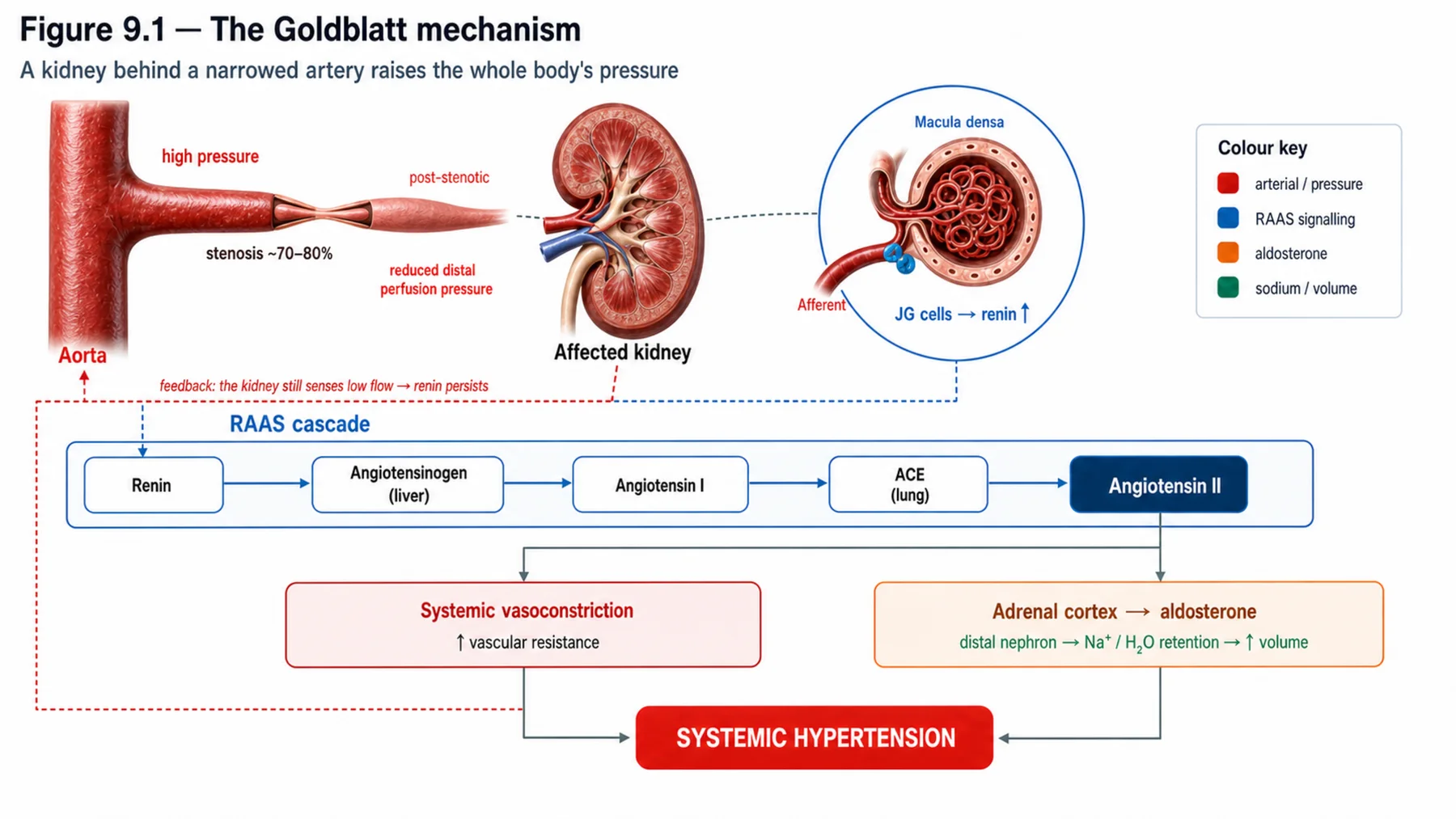
Renal artery stenosis causes hypertension by the mechanism the opening chapter foreshadowed. The narrowing reduces the perfusion pressure reaching the affected kidney, which the juxtaglomerular apparatus reads as hypotension and responds to by releasing renin; renin generates angiotensin II, which raises the systemic blood pressure by vasoconstriction and by driving aldosterone and sodium retention — the kidney, sensing it is under-perfused, raises the pressure everywhere to restore its own perfusion. This is the Goldblatt mechanism, demonstrated experimentally by clipping a renal artery. The key insight is that the hypertension is RAAS-driven, which explains both why RAAS blockade is effective and why the physiology differs so importantly between unilateral and bilateral disease. The narrowed artery is not directly raising the systemic pressure; it is doing so through the renin it provokes.
— Two diseases: atherosclerotic and fibromuscular
Renal artery stenosis comes in two forms with very different epidemiology and treatment. Atherosclerotic renal artery stenosis is by far the commoner (around 90%): it occurs in older patients with diffuse atherosclerosis, affects the proximal artery and ostium, is progressive, is frequently bilateral, and causes not only hypertension but ischaemic nephropathy — a progressive decline in kidney function from chronic hypoperfusion. Fibromuscular dysplasia is the other form: a non-atherosclerotic disease of the arterial wall affecting younger patients, characteristically women, involving the mid-to-distal artery and producing the classic 'string of beads' on angiography. The distinction matters enormously for treatment, because — as we will see — atherosclerotic disease responds to medical therapy and rarely benefits from stenting, while fibromuscular dysplasia is potentially cured by angioplasty. The patient's age, sex, and vascular risk profile usually predict which disease is present before the imaging confirms it.
— The decisive physiology: unilateral versus bilateral
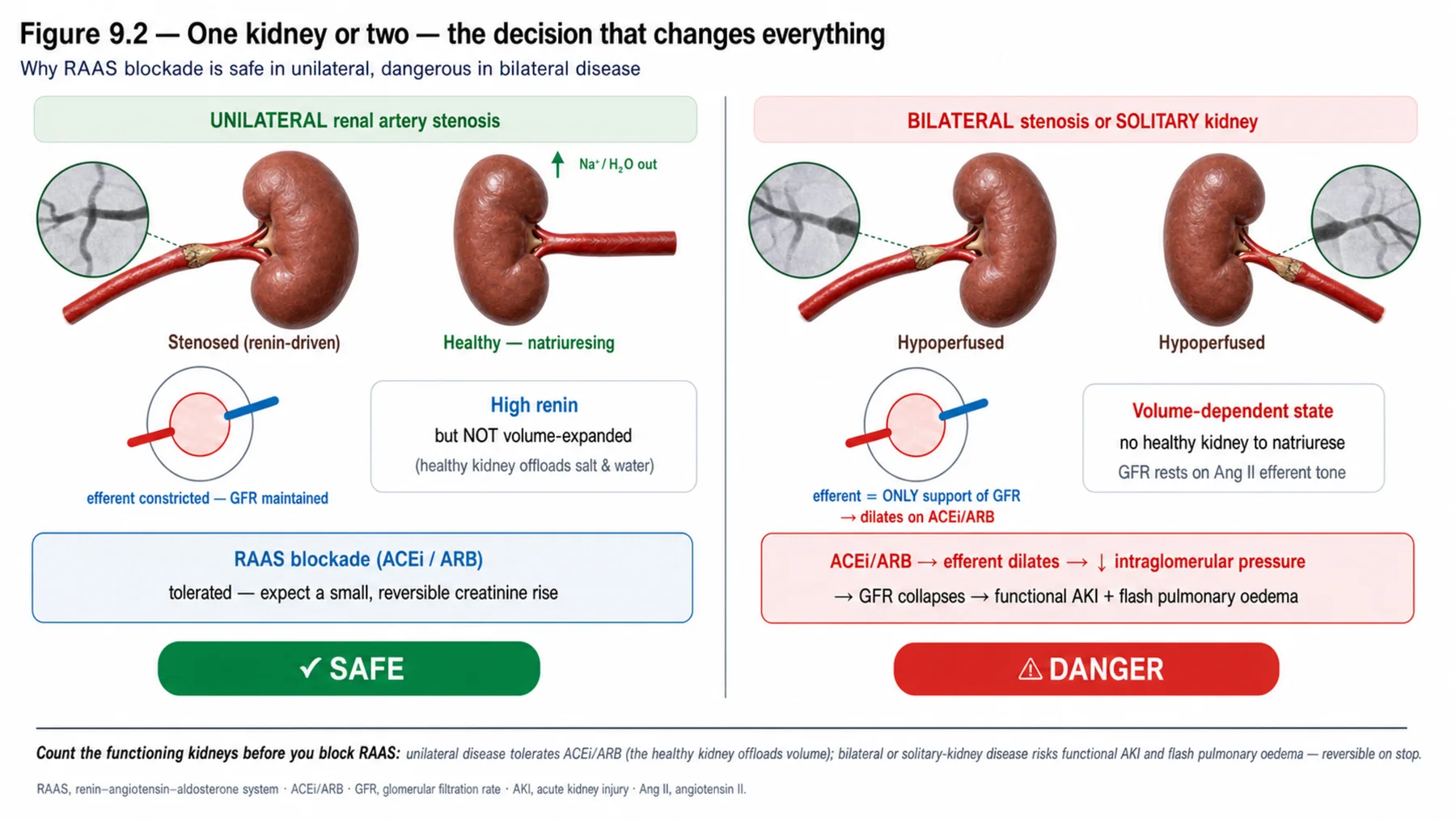
The single most important physiological distinction — and a frequent source of error — is between unilateral and bilateral stenosis. In unilateral stenosis, the affected kidney is under-perfused and drives renin and hypertension, but the contralateral, normal kidney sees the high systemic pressure and responds by natriuresis (pressure-natriuresis), excreting the sodium so that the patient is not volume-expanded; the hypertension is high-renin and not volume-dependent, and RAAS blockade is effective and safe — the affected kidney's filtration may fall, but the healthy contralateral kidney maintains overall function. In bilateral stenosis (or stenosis to a solitary functioning kidney), there is no normal kidney to natriurese: both kidneys are hypoperfused, the state becomes volume-dependent, and — critically — both kidneys depend on angiotensin-II-mediated efferent constriction to maintain their filtration. Blocking the RAAS in this setting removes that efferent support and can precipitate functional acute kidney injury, and the volume-dependence produces the characteristic recurrent 'flash' pulmonary oedema. So the same drug (a RAAS blocker) is safe and effective in unilateral disease but a hazard in bilateral disease — which is why a marked creatinine rise on starting a RAAS blocker is a classic clue to bilateral renovascular disease.
— Recognising and imaging it
The diagnostic clues follow from the mechanism and the physiology: resistant hypertension, a significant rise in creatinine after starting a RAAS blocker (pointing to bilateral disease), recurrent flash pulmonary oedema (the volume-dependent bilateral picture), an abdominal bruit, and asymmetric kidney size on imaging — with the patient profile (older vasculopath versus younger woman) pointing to the disease type. Imaging proceeds from non-invasive tests — duplex ultrasound (first-line but operator-dependent), CT angiography, and MR angiography — to catheter angiography, which is the reference standard and the route to intervention. A crucial caveat tempers all of this: atherosclerotic stenosis is common as an incidental finding in older vasculopaths, so the mere presence of a stenosis does not prove it is causing the hypertension or the renal impairment — the lesion must be plausibly functionally significant and fit the clinical picture before it is blamed and certainly before it is treated invasively. Treating an incidental stenosis is a recognised error.
— The treatment debate: medical therapy versus stenting
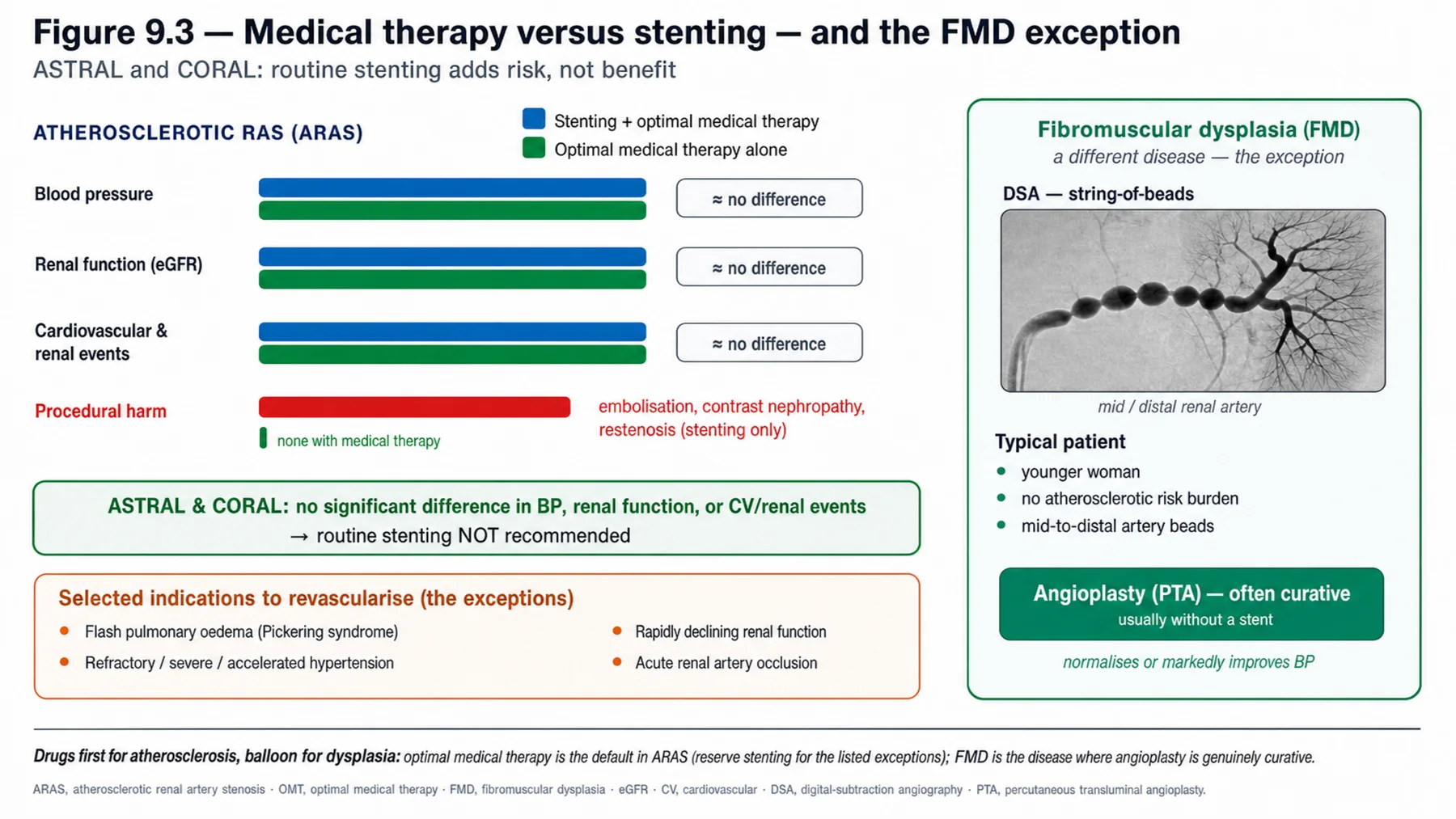
Here is the chapter's centrepiece, and one of the clearer reversals in modern nephrology. For atherosclerotic renal artery stenosis, the intuitive treatment — open the narrowed artery with a stent — was widely practised until randomised trials tested it. The major trials, ASTRAL and CORAL, compared stenting plus medical therapy against optimal medical therapy alone and found that stenting added nothing: no improvement in blood pressure, renal function, or cardiovascular events for most patients, while carrying procedural risks including atheroembolism. The explanation is that the atherosclerotic stenosis is often not the sole driver (the patient has diffuse vascular and parenchymal disease), that medical therapy addresses the RAAS and the cardiovascular risk effectively, and that opening one artery does not reverse the broader disease. So the evidence-based approach to atherosclerotic disease is optimal medical therapy first — RAAS blockade (monitored, with caution in bilateral disease), a statin, antiplatelet therapy, blood-pressure control, and aggressive cardiovascular risk reduction including smoking cessation — with stenting reserved for the minority with a clear high-risk indication: recurrent flash pulmonary oedema, rapidly or progressively declining renal function, severe or refractory hypertension despite optimal medical therapy, or acute occlusion. Routine stenting of an atherosclerotic stenosis is not beneficial.
— Fibromuscular dysplasia: the curable exception, and the synthesis
Fibromuscular dysplasia is the important exception to the medical-therapy-first rule. Because it is a non-atherosclerotic disease of a younger patient, often with a shorter duration of hypertension and without the diffuse vascular and parenchymal disease that limits the benefit of intervention in atherosclerotic stenosis, angioplasty (usually without a stent) is effective and frequently curative or substantially improving — so revascularisation is a reasonable and often preferred treatment, in contrast to the atherosclerotic form. The synthesis of the chapter is therefore a set of distinctions: recognise renal artery stenosis from its clues, classify it as atherosclerotic or fibromuscular and as unilateral or bilateral, treat atherosclerotic disease with optimal medical therapy (reserving stenting for the defined high-risk indications, because CORAL and ASTRAL showed routine stenting adds nothing), treat fibromuscular dysplasia with angioplasty, and handle RAAS blockade carefully in bilateral disease (effective for the hypertension but a risk for functional acute kidney injury). The renovascular story is, above all, a lesson that the presence of a fixable-looking lesion does not mean fixing it helps — the evidence, not the anatomy, decides.
| 04 | PHASE A · LEVEL 4 · ORIENTATION & KNOWLEDGE Reference Tables |
Table 9.1 — The Goldblatt mechanism
| Step | Detail |
| Stenosis | Reduces perfusion pressure to the affected kidney |
| Renin | Juxtaglomerular apparatus releases renin |
| Angiotensin II | Vasoconstriction + aldosterone + sodium retention → systemic hypertension |
| Implication | RAAS-driven — explains RAAS-blockade efficacy and the unilateral/bilateral difference |
Table 9.2 — Atherosclerotic vs fibromuscular
| Feature | Atherosclerotic (ARAS) | Fibromuscular dysplasia (FMD) |
| Frequency / patient | ~90%; older vasculopath | Younger, often women |
| Site | Proximal/ostial; progressive; often bilateral | Mid-to-distal; 'string of beads' |
| Consequence | Hypertension + ischaemic nephropathy | Hypertension (potentially curable) |
| Treatment | Medical therapy first; selective stenting | Angioplasty — often curative |
Table 9.3 — Unilateral versus bilateral physiology
| Unilateral | Bilateral / solitary kidney | |
| Renin/volume | High-renin; not volume-expanded | Volume-dependent; both kidneys hypoperfused |
| Contralateral kidney | Natriureses (pressure-natriuresis) | No normal kidney to compensate |
| RAAS blockade | Effective and safe | Risk of functional AKI; flash pulmonary oedema |
Table 9.4 — Diagnostic clues and imaging
| Item | Detail |
| Clues | Resistant HTN, Cr rise on RAAS blockade, flash pulmonary oedema, abdominal bruit, asymmetric kidneys |
| Imaging (non-invasive) | Duplex ultrasound (first-line), CT angiography, MR angiography |
| Reference standard | Catheter angiography (and route to intervention) |
| Caveat | Incidental ARAS is common — a stenosis is not always the cause |
Table 9.5 — Treatment of atherosclerotic disease
| Element | Detail |
| Medical therapy (first) | RAAS blockade (monitored), statin, antiplatelet, BP/CV risk control, smoking cessation |
| Stenting evidence | ASTRAL, CORAL — no benefit over medical therapy for most |
| Revascularise (selected) | Recurrent flash pulmonary oedema; rapidly declining renal function; refractory/severe HTN; acute occlusion |
| Routine stenting | Not beneficial — procedural risk (atheroembolism) |
Table 9.6 — The evidence and the synthesis
| Point | Detail |
| ARAS | Medical therapy first; stenting no routine benefit (CORAL/ASTRAL) |
| FMD | Angioplasty — often curative; revascularise |
| RAAS blockade | Safe/effective unilateral; AKI risk in bilateral (monitor) |
| The lesson | A fixable-looking lesion does not mean fixing it helps — the evidence decides |
| 05 | PHASE B · LEVEL 5 · VISUALISE & MAP Imaging & Flowchart Specifications |
| 06 | PHASE B · LEVEL 6 · VISUALISE & MAP Concept Maps |
Each chain runs from mechanism to a named bedside action; read the arrows as “leads to.”
Goldblatt mechanism. Renal artery stenosis → ↓ perfusion pressure → renin → angiotensin II → systemic hypertension → ACTION: recognise RAAS-driven hypertension and use RAAS blockade (with the unilateral/bilateral caveat).
Unilateral vs bilateral. Unilateral (contralateral kidney natriureses; high-renin, RAAS blockade safe) vs bilateral/solitary (volume-dependent; both kidneys depend on angiotensin II) → ACTION: use RAAS blockade confidently in unilateral disease but monitor for functional AKI in bilateral.
ARAS vs FMD. Atherosclerotic (older, proximal, ischaemic nephropathy) vs fibromuscular (younger women, distal, string of beads) → ACTION: classify the disease — medical therapy for atherosclerotic, angioplasty for fibromuscular.
The stenting evidence. Atherosclerotic stenosis is often not the sole driver + diffuse disease → stenting adds nothing (CORAL/ASTRAL) → ACTION: treat medically first; reserve stenting for defined high-risk indications.
RAAS blockade risk in bilateral. Both kidneys depend on angiotensin-II efferent tone → RAAS blockade removes it → functional AKI → ACTION: a marked creatinine rise on starting a RAAS blocker is a clue to bilateral disease — monitor and reconsider.
| 07 | PHASE B · LEVEL 7 · VISUALISE & MAP Decision Pathways |
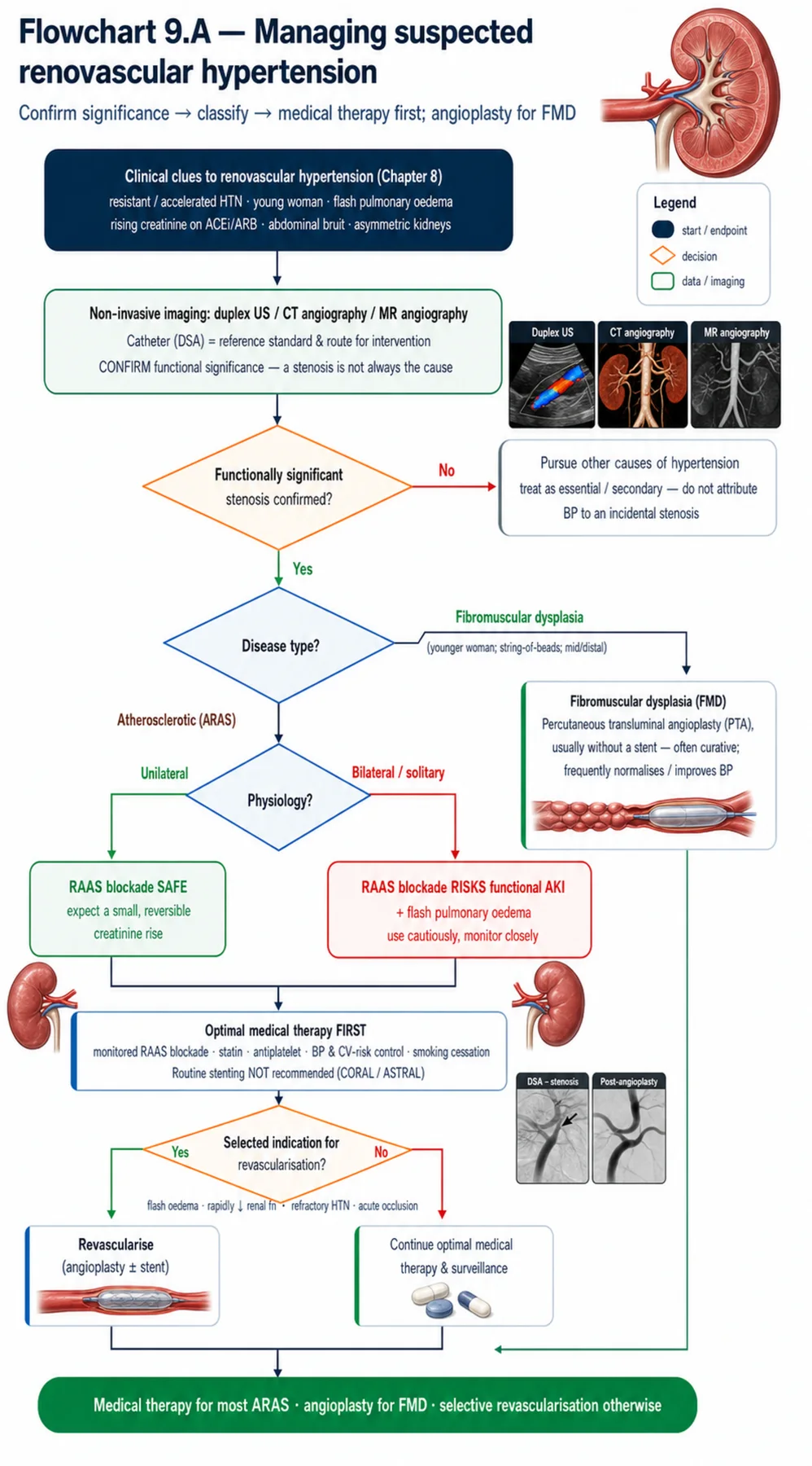
| R1 | IF renovascular hypertension is suspected, THEN recognise it as RAAS-driven (the Goldblatt mechanism) and image the renal arteries. |
| R2 | IF a stenosis is found, THEN confirm it is functionally significant and fits the clinical picture — an incidental stenosis is not always the cause. |
| R3 | IF the patient is an older vasculopath, THEN expect atherosclerotic disease; IF a younger woman with a string-of-beads appearance, THEN expect fibromuscular dysplasia. |
| R4 | IF the disease is unilateral, THEN use RAAS blockade confidently; IF bilateral or to a solitary kidney, THEN monitor closely for functional acute kidney injury. |
| R5 | IF the disease is atherosclerotic, THEN treat with optimal medical therapy first — stenting adds nothing for most patients (CORAL, ASTRAL). |
| R6 | IF there is recurrent flash pulmonary oedema, rapidly declining renal function, or refractory/severe hypertension despite optimal medical therapy, THEN consider revascularisation. |
| R7 | IF the disease is fibromuscular dysplasia, THEN consider angioplasty — it is often curative. |
| R8 | IF a marked creatinine rise follows starting a RAAS blocker, THEN suspect bilateral renovascular disease. |
| 08 | PHASE C · LEVEL 8 · CLINICAL REASONING Clinical Cases |
| CASE 1 | DON'T STENT IT Medical therapy first Atherosclerotic renal artery stenosis |
Presentation
An older patient with diffuse atherosclerosis has hypertension and an incidentally found atherosclerotic renal artery stenosis. An interventionalist proposes stenting the artery to cure the hypertension.
❖ Pause and reflect Will stenting the atherosclerotic stenosis improve this patient's outcome? |
Analysis
Probably not — the major trials say otherwise. ASTRAL and CORAL compared stenting plus medical therapy against optimal medical therapy alone for atherosclerotic renal artery stenosis and found no improvement in blood pressure, renal function, or cardiovascular events from stenting, with procedural risk (including atheroembolism). In this older vasculopath, the stenosis is often not the sole driver, the disease is diffuse, and medical therapy addresses the RAAS and cardiovascular risk effectively. The evidence-based approach is optimal medical therapy first — not stenting — reserving revascularisation for specific high-risk presentations this patient does not have.
Plan
Treat with optimal medical therapy — RAAS blockade (monitored), a statin, antiplatelet therapy, blood-pressure and cardiovascular risk control, and smoking cessation — rather than stenting the atherosclerotic stenosis, per CORAL and ASTRAL. Reserve revascularisation for defined indications.
Teaching point
For atherosclerotic renal artery stenosis, optimal medical therapy comes first — routine stenting adds nothing (CORAL, ASTRAL).
Cross-reference
Exercises rules R2 and R5; the stenting-evidence concept map; Figure 9.3; Tables 9.5, 9.6.
| CASE 2 | AKI ON THE ACE INHIBITOR The bilateral physiology Bilateral renovascular disease |
Presentation
A patient with hypertension develops a marked rise in creatinine and recurrent flash pulmonary oedema shortly after starting an ACE inhibitor. Imaging shows bilateral renal artery stenosis.
❖ Pause and reflect Why did the ACE inhibitor precipitate AKI and flash oedema here? |
Analysis
Because of the bilateral physiology. With both renal arteries stenosed (and no normal kidney to compensate), both kidneys are hypoperfused and depend on angiotensin-II-mediated efferent constriction to maintain their filtration; the ACE inhibitor removes that efferent support and precipitates functional acute kidney injury — hence the marked creatinine rise. The bilateral disease is also volume-dependent, producing the recurrent flash pulmonary oedema. This is the classic presentation that distinguishes bilateral from unilateral disease: a marked creatinine rise on starting a RAAS blocker is a clue to bilateral renovascular disease, and the flash pulmonary oedema is itself an indication to consider revascularisation.
Plan
Recognise the bilateral renovascular disease, manage the RAAS blockade carefully (monitor; a marked rise may force reconsideration), and — given the recurrent flash pulmonary oedema — consider revascularisation, which is one of the selected indications. Handle RAAS blockade cautiously in bilateral disease.
Teaching point
In bilateral renovascular disease, RAAS blockade can precipitate functional AKI, and recurrent flash pulmonary oedema is characteristic and an indication to revascularise.
Cross-reference
Exercises rules R4, R6, R8; the unilateral-versus-bilateral and RAAS-risk concept maps; Figure 9.2; Tables 9.3, 9.5.
| CASE 3 | THE STRING OF BEADS Angioplasty can cure Fibromuscular dysplasia |
Presentation
A young woman with recent-onset hypertension has a 'string of beads' appearance in the mid-to-distal renal artery on angiography. The team, recalling that stenting renal arteries 'doesn't work,' plans medical therapy alone.
❖ Pause and reflect Does the 'stenting doesn't work' lesson apply to this fibromuscular dysplasia? |
Analysis
No — fibromuscular dysplasia is the important exception. The 'stenting adds nothing' evidence (CORAL, ASTRAL) applies to atherosclerotic renal artery stenosis, not to fibromuscular dysplasia. This young woman with a string-of-beads appearance has fibromuscular dysplasia, a non-atherosclerotic disease without the diffuse vascular and parenchymal disease that limits the benefit of intervention in the atherosclerotic form; angioplasty (usually without a stent) is effective and frequently curative, especially given her young age and short duration of hypertension. Treating her with medical therapy alone, on the mistaken extension of the atherosclerotic evidence, would forgo a potential cure.
Plan
Consider angioplasty for the fibromuscular dysplasia — it is often curative in a young patient with short-duration hypertension — rather than applying the atherosclerotic 'stenting doesn't work' evidence. Revascularise fibromuscular dysplasia.
Teaching point
Fibromuscular dysplasia (young woman, string of beads) is often cured by angioplasty — the atherosclerotic 'stenting adds nothing' evidence does not apply.
Cross-reference
Exercises rules R3 and R7; the ARAS-vs-FMD concept map; Figure 9.3; Tables 9.2, 9.6.
| CASE 4 | WHEN TO REVASCULARISE The selected indication High-risk atherosclerotic presentation |
Presentation
An older patient with atherosclerotic bilateral renal artery stenosis has rapidly declining renal function and refractory hypertension despite genuinely optimal medical therapy. A clinician, having learned that 'stenting doesn't work,' is reluctant to revascularise.
❖ Pause and reflect Is this a patient in whom revascularisation should be considered despite the general evidence? |
Analysis
Yes — this is one of the selected indications. The CORAL/ASTRAL evidence shows that routine stenting of atherosclerotic stenosis adds nothing, but those trials largely excluded or under-represented the high-risk presentations in which revascularisation may still help: recurrent flash pulmonary oedema, rapidly or progressively declining renal function, severe or refractory hypertension despite genuinely optimal medical therapy, and acute occlusion. This patient has rapidly declining function and refractory hypertension on optimal therapy, so revascularisation should be considered despite the general 'medical therapy first' rule. The lesson is nuance: routine stenting is not beneficial, but defined high-risk presentations are the exception.
Plan
Consider revascularisation given the rapidly declining renal function and refractory hypertension despite optimal medical therapy — a recognised selected indication — rather than withholding it on the general evidence. Revascularise the defined high-risk atherosclerotic presentations.
Teaching point
Revascularisation of atherosclerotic disease is reserved for selected high-risk presentations — flash pulmonary oedema, rapidly declining function, refractory/severe HTN — despite the general 'medical therapy first' rule.
Cross-reference
Exercises rule R6; the stenting-evidence concept map; Table 9.5.
| 09 | PHASE C · LEVEL 9 · CLINICAL REASONING Clinical Implications |
One triad per mechanism the narrative exposed: the physiology, why it matters, and the bedside move.
MECHANISM Renal artery stenosis reduces perfusion pressure and activates the RAAS (Goldblatt). |
WHY IT MATTERS The hypertension is RAAS-driven, which explains the response to RAAS blockade. |
ACTION Recognise renovascular hypertension as RAAS-driven and image the renal arteries. |
MECHANISM In unilateral disease the contralateral kidney natriureses; in bilateral disease neither can. |
WHY IT MATTERS RAAS blockade is safe in unilateral but risks functional AKI in bilateral disease. |
ACTION Use RAAS blockade confidently in unilateral disease; monitor closely in bilateral disease. |
MECHANISM Atherosclerotic and fibromuscular disease differ in patient, anatomy, and treatment. |
WHY IT MATTERS Medical therapy suits atherosclerotic disease; angioplasty suits fibromuscular dysplasia. |
ACTION Classify the disease before deciding on revascularisation. |
MECHANISM Atherosclerotic stenosis is often not the sole driver, amid diffuse disease. |
WHY IT MATTERS Stenting it adds nothing to medical therapy for most patients (CORAL, ASTRAL). |
ACTION Treat atherosclerotic disease medically first; reserve stenting for defined indications. |
MECHANISM Both kidneys in bilateral disease depend on angiotensin-II efferent tone. |
WHY IT MATTERS A marked creatinine rise on a RAAS blocker signals bilateral disease. |
ACTION Suspect bilateral renovascular disease when the creatinine climbs on starting a RAAS blocker. |
| 10 | PHASE C · LEVEL 10 · CLINICAL REASONING Clinical Pearls |
| Renal artery stenosis → ↓ perfusion → renin → angiotensin II → systemic HTN (Goldblatt). | ARAS ~90%: older vasculopath, proximal/ostial, progressive, often bilateral, ischaemic nephropathy. |
| FMD: younger women, mid-distal, 'string of beads', potentially curable. | Unilateral: high-renin, contralateral natriureses, RAAS blockade safe. |
| Bilateral/solitary: volume-dependent, both depend on angiotensin II, RAAS blockade → functional AKI. | Marked Cr rise on starting a RAAS blocker → suspect bilateral disease. |
| Flash (recurrent) pulmonary oedema → bilateral renovascular disease. | Clues: resistant HTN, Cr rise on RAAS blockade, bruit, asymmetric kidneys. |
| Imaging: duplex (first-line), CTA, MRA; catheter angiography reference. | Incidental ARAS is common — a stenosis is not always the cause. |
| ARAS: optimal medical therapy FIRST (RAAS blockade, statin, antiplatelet, BP/CV risk). | Stenting ARAS: NO routine benefit (CORAL, ASTRAL) + procedural risk. |
| Revascularise ARAS for: flash oedema, rapidly declining function, refractory/severe HTN, acute occlusion. | FMD: angioplasty — often curative; revascularise. |
| RAAS blockade in renovascular: effective; monitor in bilateral. | A fixable-looking lesion doesn't mean fixing it helps — the evidence decides. |
| 11 | PHASE D · LEVEL 11 · SAFETY & EVIDENCE Red Flags & Never-Do |
Panel A — Red flags
| ▲ | A marked creatinine rise on starting an ACE inhibitor or ARB — suspect bilateral renovascular disease. |
| ▲ | Recurrent flash pulmonary oedema — bilateral renovascular disease; an indication to revascularise. |
| ▲ | An incidental atherosclerotic stenosis being blamed for the hypertension — a stenosis is not always the cause. |
| ▲ | A young woman with a string-of-beads appearance — fibromuscular dysplasia; angioplasty can cure. |
| ▲ | Rapidly declining renal function or refractory HTN despite optimal medical therapy — consider revascularisation. |
Panel B — Never do
| ✖ NEVER — routinely stent an atherosclerotic renal artery stenosis (CORAL, ASTRAL). |
| ✖ NEVER — apply the atherosclerotic 'stenting adds nothing' evidence to fibromuscular dysplasia. |
| ✖ NEVER — blame and treat an incidental stenosis without confirming functional significance. |
| ✖ NEVER — start a RAAS blocker in suspected bilateral disease without monitoring renal function. |
| 12 | PHASE D · LEVEL 12 · SAFETY & EVIDENCE Common Pitfalls |
Pitfall 1 — Stenting atherosclerotic disease
| ✖ | WRONG Stenting an atherosclerotic renal artery stenosis to 'cure' the hypertension. |
| ✓ | RIGHT Treating with optimal medical therapy first. |
| ✉ | WHY Stenting adds nothing to medical therapy for most (CORAL, ASTRAL). |
Pitfall 2 — Withholding angioplasty in FMD
| ✖ | WRONG Treating fibromuscular dysplasia medically because 'stenting doesn't work.' |
| ✓ | RIGHT Offering angioplasty, which is often curative. |
| ✉ | WHY The atherosclerotic evidence does not apply to fibromuscular dysplasia. |
Pitfall 3 — Blaming an incidental stenosis
| ✖ | WRONG Attributing hypertension to an incidentally found stenosis. |
| ✓ | RIGHT Confirming functional significance and clinical fit first. |
| ✉ | WHY Incidental atherosclerotic stenosis is common and often not the cause. |
Pitfall 4 — RAAS blockade without monitoring in bilateral disease
| ✖ | WRONG Starting a RAAS blocker in bilateral disease without checking renal function. |
| ✓ | RIGHT Monitoring closely for a functional creatinine rise. |
| ✉ | WHY Both kidneys depend on angiotensin-II efferent tone. |
Pitfall 5 — Missing the selected indication
| ✖ | WRONG Withholding revascularisation in flash oedema or rapidly declining function. |
| ✓ | RIGHT Recognising these as selected indications to revascularise. |
| ✉ | WHY Routine stenting is futile, but defined high-risk presentations are the exception. |
| 13 | PHASE D · LEVEL 13 · SAFETY & EVIDENCE Evidence Grading |
GRADE A HIGH CONFIDENCE The effect is real and the estimate is stable. RCTs at low risk of bias; multiple concordant prospective cohorts; meta-analyses. |
GRADE B MODERATE CONFIDENCE The effect is likely real but may shift with new data. Observational studies, registries, mechanistic human studies. |
GRADE C LOW CONFIDENCE Rests on physiology, reasoning, or consensus rather than outcomes. Pathophysiological reasoning; extrapolation; consensus without outcomes. |
Graded statements (by evidence type)
| Statement | Grade | Basis (evidence type) |
| Renal artery stenosis causes hypertension via RAAS activation (Goldblatt). | A | Established physiology |
| Stenting atherosclerotic stenosis adds nothing to medical therapy for most. | A | RCTs (ASTRAL, CORAL) |
| Optimal medical therapy is first-line for atherosclerotic disease. | A | RCTs and consensus |
| Bilateral disease risks functional AKI on RAAS blockade. | A | Established physiology |
| Selected presentations may benefit from revascularisation. | B | Observational and subgroup data |
| Angioplasty is effective and often curative in fibromuscular dysplasia. | B | Observational data |
| Incidental atherosclerotic stenosis is common and often not causal. | A | Epidemiological data |
| 14 | PHASE E · LEVEL 14 · PATIENT DECISIONS Absolute Risk in Natural Frequency |
Natural-frequency estimates for orientation, from the renovascular trials; they vary with the population. They convey the size of the decisions, expressed per 100 comparable patients.
| Per 100 patients… | Outcome | Roughly how many | See |
| Atherosclerotic disease stented vs medical therapy | Gain a benefit in BP/renal/CV outcomes | No more — stenting adds nothing | L13 row 2 |
| Atherosclerotic disease stented | Suffer a procedural complication | Some — hence the harm side | L13 row 2 |
| Fibromuscular dysplasia treated with angioplasty | Be cured or substantially improved | Many | L13 row 6 |
| Bilateral disease started on a RAAS blocker unmonitored | Develop functional AKI | More than the monitored | L13 row 4 |
★ How to read these Read these as orientation, not promises; outcomes vary with the population. The stable signals: stenting atherosclerotic disease adds nothing (and carries procedural risk), angioplasty helps fibromuscular dysplasia, and bilateral disease risks AKI on RAAS blockade. Communicate them as people out of 100, not as a hazard ratio. |
| 17 | PHASE F · LEVEL 17 · APPLY & TEST Documentation Templates |
Paste-ready notes. Tick the boxes that apply and delete the rest; make the classification and the evidence-based decision explicit.
Template 1 — Renovascular work-up and medical therapy
Template 2 — Revascularisation decision
| 18 | PHASE F · LEVEL 18 · APPLY & TEST Cheat Sheet |
| RAS → ↓ perfusion → renin → angiotensin II → HTN (Goldblatt). | ARAS ~90%: older, proximal, progressive, often bilateral, ischaemic nephropathy. |
| FMD: younger women, mid-distal, string of beads, curable. | Unilateral: high-renin, contralateral natriureses, RAAS blockade safe. |
| Bilateral/solitary: volume-dependent, RAAS blockade → functional AKI. | Marked Cr rise on RAAS blocker → bilateral disease. |
| Flash (recurrent) pulmonary oedema → bilateral. | Clues: resistant HTN, Cr rise on RAAS blockade, bruit, asymmetric kidneys. |
| Imaging: duplex → CTA/MRA → catheter angiography. | Incidental ARAS common — stenosis ≠ cause. |
| ARAS: medical therapy FIRST. | Stenting ARAS: NO routine benefit (CORAL, ASTRAL). |
| Revascularise ARAS: flash oedema, rapidly declining function, refractory HTN, acute occlusion. | FMD: angioplasty — often curative. |
| RAAS blockade: monitor in bilateral disease. | Fixable-looking lesion ≠ fixing it helps — evidence decides. |
| 19 | PHASE F · LEVEL 19 · APPLY & TEST Flashcards |
| CARD 1 | Q. What is the Goldblatt mechanism? A. Renal artery stenosis reduces perfusion pressure to the affected kidney, which the juxtaglomerular apparatus reads as hypotension and responds to by releasing renin; the resulting angiotensin II raises systemic blood pressure by vasoconstriction and sodium retention. DETAILED. The hypertension is RAAS-driven. CLINICAL. Recognise renovascular hypertension as RAAS-driven and image the renal arteries. |
| CARD 2 | Q. How do atherosclerotic and fibromuscular renal artery disease differ? A. Atherosclerotic stenosis (~90%) affects older vasculopaths, is proximal/ostial, progressive, often bilateral, and causes ischaemic nephropathy; fibromuscular dysplasia affects younger women, involves the mid-to-distal artery (string of beads), and is potentially curable by angioplasty. DETAILED. They need different treatments. CLINICAL. Classify the disease before deciding on revascularisation. |
| CARD 3 | Q. Why does the unilateral/bilateral distinction matter for RAAS blockade? A. In unilateral disease the contralateral kidney natriureses, so the patient is not volume-expanded and RAAS blockade is safe; in bilateral disease (or a solitary kidney) both kidneys depend on angiotensin-II efferent tone to filter, so RAAS blockade can precipitate functional acute kidney injury. DETAILED. A marked creatinine rise on a RAAS blocker suggests bilateral disease. CLINICAL. Use RAAS blockade confidently in unilateral disease but monitor in bilateral. |
| CARD 4 | Q. What do ASTRAL and CORAL show about stenting? A. That stenting atherosclerotic renal artery stenosis adds nothing to optimal medical therapy for blood pressure, renal function, or cardiovascular events in most patients, while carrying procedural risk. DETAILED. The stenosis is often not the sole driver amid diffuse disease. CLINICAL. Treat atherosclerotic disease medically first; do not routinely stent. |
| CARD 5 | Q. What is the treatment of atherosclerotic renal artery stenosis? A. Optimal medical therapy first — RAAS blockade (monitored), a statin, antiplatelet therapy, blood-pressure and cardiovascular risk control, and smoking cessation — with revascularisation reserved for selected high-risk presentations. DETAILED. Routine stenting is not beneficial. CLINICAL. Optimise medical therapy; revascularise only for defined indications. |
| CARD 6 | Q. When should atherosclerotic disease be revascularised? A. For recurrent flash pulmonary oedema, rapidly or progressively declining renal function, severe or refractory hypertension despite genuinely optimal medical therapy, and acute occlusion — the selected high-risk presentations. DETAILED. Routine stenting is futile, but these are the exceptions. CLINICAL. Revascularise the defined high-risk atherosclerotic presentations. |
| CARD 7 | Q. How is fibromuscular dysplasia treated? A. With angioplasty (usually without a stent), which is effective and frequently curative, especially in younger patients with a shorter duration of hypertension — the atherosclerotic 'stenting adds nothing' evidence does not apply. DETAILED. It is the curable exception. CLINICAL. Offer angioplasty for fibromuscular dysplasia. |
| CARD 8 | Q. Why is an incidental stenosis a trap? A. Atherosclerotic stenosis is common as an incidental finding in older vasculopaths, so a stenosis does not prove it is causing the hypertension or renal impairment — it must be functionally significant and fit the clinical picture before being blamed or treated. DETAILED. Treating an incidental stenosis is a recognised error. CLINICAL. Confirm functional significance before attributing or intervening. |
| 20 | PHASE F · LEVEL 20 · APPLY & TEST One-Minute Preceptor |
| SCENE 1 | The interventionalist keen to stent |
GET A COMMITMENT. “You want to stent this older patient's atherosclerotic renal artery stenosis to cure the hypertension — why?”
PROBE FOR EVIDENCE. “Opening the artery should fix it” — ask: “What did ASTRAL and CORAL show about stenting versus medical therapy?”
TEACH A GENERAL RULE. For atherosclerotic stenosis, stenting adds nothing to optimal medical therapy in most patients, with procedural risk — the stenosis is often not the sole driver amid diffuse disease.
REINFORCE WHAT WAS RIGHT. Recognising the stenosis was correct.
CORRECT A MISTAKE. Optimise medical therapy; reserve stenting for defined high-risk indications.
| SCENE 2 | The resident over-applying the evidence |
GET A COMMITMENT. “You're treating this young woman's string-of-beads stenosis medically because 'stenting doesn't work' — is that right here?”
PROBE FOR EVIDENCE. “The trials said no benefit” — ask: “Did CORAL and ASTRAL study fibromuscular dysplasia, and what does angioplasty do for it?”
TEACH A GENERAL RULE. The 'stenting adds nothing' evidence is for atherosclerotic disease; fibromuscular dysplasia is often cured by angioplasty, so the rule doesn't apply.
REINFORCE WHAT WAS RIGHT. Knowing the atherosclerotic evidence was good.
CORRECT A MISTAKE. Offer angioplasty for the fibromuscular dysplasia.
| 21 | PHASE F · LEVEL 21 · APPLY & TEST Reflective Prompts |
Genuine tensions this evidence leaves open; sit with them rather than resolving them too quickly.
A narrowed artery and a clear mechanism made stenting feel obviously right — yet the trials said no. How do you weigh mechanistic plausibility against outcome evidence when they conflict?
The trials excluded the high-risk presentations in which intervention might still help, leaving a grey zone. How do you act in the patients the evidence did not study?
Incidental stenosis is common, and the temptation to treat what you can see is strong. How do you resist intervening on an anatomical finding that may be a bystander?
The same lesion (a renal artery narrowing) is left alone in one patient (atherosclerotic) and opened in another (fibromuscular). How comfortable are you treating identical-looking anatomy oppositely on the basis of disease type?
RAAS blockade is the right drug and a potential hazard in the same disease, depending on whether one or both arteries are stenosed. How do you hold that dual nature in mind at the bedside?
| 22 | PHASE F · LEVEL 22 · APPLY & TEST Board-Style Questions |
| Q 01 | Renal artery stenosis causes hypertension by: |
| A | Directly raising systemic vascular resistance |
| B | Reducing renal perfusion, activating renin and angiotensin II (Goldblatt) |
| C | Causing volume depletion |
| D | Blocking aldosterone |
Rationale The stenosis reduces perfusion, triggering renin and angiotensin II — the Goldblatt mechanism (Figure 9.1, rule R1). A, C, and D are incorrect. |
| Q 02 | What do ASTRAL and CORAL show about stenting atherosclerotic renal artery stenosis? |
| A | It cures the hypertension |
| B | It adds nothing to optimal medical therapy for most patients |
| C | It is always harmful |
| D | It improves renal function dramatically |
Rationale Stenting added no benefit over medical therapy for most patients, with procedural risk (case 1, Figure 9.3, rule R5). A and D overstate; C is too absolute. |
| Q 03 | Why can a RAAS blocker precipitate acute kidney injury in bilateral renal artery stenosis? |
| A | It causes volume overload |
| B | Both kidneys depend on angiotensin-II efferent constriction to maintain filtration |
| C | It is directly nephrotoxic |
| D | It raises the blood pressure |
Rationale In bilateral disease both kidneys rely on angiotensin-II efferent tone, which the blocker removes (case 2, Figure 9.2, rule R4). A, C, and D are incorrect. |
| Q 04 | A young woman with hypertension and a 'string of beads' renal artery has: |
| A | Atherosclerotic stenosis — treat medically |
| B | Fibromuscular dysplasia — consider angioplasty (often curative) |
| C | An incidental finding to ignore |
| D | Bilateral atherosclerosis |
Rationale The string of beads in a young woman is fibromuscular dysplasia, treated with angioplasty (case 3, Table 9.2, rule R7). A misapplies the atherosclerotic evidence; C and D are wrong. |
| Q 05 | Which is a recognised indication to revascularise atherosclerotic disease? |
| A | An incidental stenosis |
| B | Recurrent flash pulmonary oedema or rapidly declining renal function despite optimal medical therapy |
| C | Mild controlled hypertension |
| D | Unilateral disease responding to medical therapy |
Rationale These selected high-risk presentations are the exception to medical-therapy-first (case 4, Table 9.5, rule R6). A, C, and D do not warrant revascularisation. |
| Q 06 | A marked creatinine rise on starting an ACE inhibitor should raise suspicion of: |
| A | Unilateral renal artery stenosis |
| B | Bilateral renal artery stenosis (or a solitary kidney) |
| C | Fibromuscular dysplasia cured |
| D | Primary aldosteronism |
Rationale Bilateral disease (both kidneys angiotensin-II-dependent) is suggested by a marked creatinine rise (case 2, rule R8). A is usually tolerated; C and D are unrelated. |
| Q 07 | First-line treatment of atherosclerotic renal artery stenosis is: |
| A | Stenting |
| B | Optimal medical therapy (RAAS blockade, statin, antiplatelet, risk control) |
| C | Surgery |
| D | Nephrectomy |
Rationale Optimal medical therapy is first-line; routine stenting is not beneficial (Table 9.5, rule R5). A, C, and D are not first-line. |
| Q 08 | An incidentally found atherosclerotic renal artery stenosis: |
| A | Always causes the hypertension |
| B | Is not necessarily the cause — confirm functional significance |
| C | Should always be stented |
| D | Excludes primary hypertension |
Rationale Incidental stenosis is common and often not causal, so functional significance must be confirmed (rule R2, Table 9.4). A, C, and D are incorrect. |