Chapter Preamble
Signals declared
Sig-D — Diagnostic (primary). Use the clinical setting to predict the characteristic disturbance pattern — the critical-care, gastrointestinal, endocrine, and refeeding signatures.
Sig-T — Therapeutic (strong). Treat the setting and correct the specific disturbances, anticipating the predictable ones (refeeding, de-resuscitation).
Sig-M — Mechanistic (strong). Why each setting generates its pattern — the acid lost in upper versus lower gut, the hormone deficiencies of adrenal disease, the insulin surge of refeeding.
Levels populated and omitted
Populated (19): L1–L14, L17–L20, L22. The mechanistic signal fires the concept maps (L6) and triads (L9); the therapeutic signal fires the absolute-risk table (L14) and templates (L17); the diagnostic signal drives the tables, rules, cases, pitfalls, and board items.
L15 / L16 preference-sensitive map and SDM scripts — omitted. No Sig-E; managing these disturbances is effective care.
L21 reflective prompts — omitted. No Sig-E/V; the chapter's tensions (the setting-predicts-pattern shortcut) are worked through the cases and pitfalls.
| 01 | PHASE A · LEVEL 1 · ORIENTATION & KNOWLEDGE Learning Objectives |
By the end of this chapter you should be able to:
Explain why specific clinical settings produce characteristic disturbance patterns.
Recognise the critical-care patterns — lactic acidosis, mixed disorders, hyperchloraemic acidosis, and overload.
Contrast upper and lower gastrointestinal losses and their opposite acid-base effects.
Recognise the endocrine patterns — adrenal insufficiency, mineralocorticoid excess, and the diabetic disturbances.
Recognise, prevent, and treat refeeding syndrome.
Use the setting as a diagnostic shortcut, then confirm and treat.
Treat the underlying setting alongside correcting the specific disturbances.
Anticipate the predictable disturbances of each setting.
| 02 | PHASE A · LEVEL 2 · ORIENTATION & KNOWLEDGE Executive Summary |
Specific clinical settings generate predictable, recognisable patterns of fluid, electrolyte, and acid-base disturbance, because the underlying pathophysiology dictates them.
Recognising the setting is therefore a powerful diagnostic shortcut: predict the pattern, then confirm and treat.
In critical care, lactic acidosis from hypoperfusion is common, often as part of a mixed disorder, alongside hyperchloraemic acidosis from saline, the acid-base and electrolyte consequences of acute kidney injury, and fluid overload.
Critical-care disturbances are frequently mixed, so the systematic and mixed-disorder methods apply directly.
Upper gastrointestinal losses (vomiting, nasogastric suction) lose hydrochloric acid, producing a metabolic alkalosis with hypokalaemia, hypochloraemia, and volume depletion — saline-responsive.
Lower gastrointestinal losses (diarrhoea, fistulae, ileostomy) lose bicarbonate-rich fluid, producing a normal-anion-gap metabolic acidosis with hypokalaemia and volume depletion.
The contrast is the key: upper gut causes alkalosis, lower gut causes acidosis, and both cause hypokalaemia and volume depletion.
Adrenal insufficiency, from aldosterone and cortisol deficiency, causes hyponatraemia, hyperkalaemia, and a metabolic acidosis — a recognisable triad.
Mineralocorticoid excess causes the opposite: hypokalaemia, metabolic alkalosis, and hypertension.
The diabetic disturbances include diabetic ketoacidosis (high-gap acidosis with potassium shifts), the hyperosmolar state, diabetes insipidus, and the type 4 renal tubular acidosis of hyporeninaemic hypoaldosteronism.
Refeeding syndrome occurs when the malnourished or alcoholic patient is fed: an insulin surge drives phosphate, potassium, and magnesium into cells, with thiamine deficiency, risking cardiac, respiratory, and neurological harm.
Refeeding hypophosphataemia is the hallmark, and the syndrome is prevented by identifying high-risk patients, feeding cautiously, replacing phosphate, potassium, and magnesium, and giving thiamine.
Across all settings, the principle is to treat the underlying setting, correct the specific disturbances by their own rules, and anticipate the predictable ones.
| 03 | PHASE A · LEVEL 3 · ORIENTATION & KNOWLEDGE Main Narrative |
Some of the most useful knowledge in this field is pattern recognition: a clinical setting predicts the disturbance. The vomiting patient will be alkalotic and hypokalaemic; the diarrhoeal patient acidotic and hypokalaemic; the patient with adrenal insufficiency hyponatraemic, hyperkalaemic, and acidotic; the refed malnourished patient hypophosphataemic. Knowing these signatures turns the diagnostic process around — the setting tells you what to look for, the numbers confirm it. This chapter assembles the characteristic patterns of four settings where these disturbances cluster.
— Critical care: lactate, mixtures, and overload
The intensive care unit concentrates fluid, electrolyte, and acid-base disturbances, and they are usually mixed. Lactic acidosis from hypoperfusion (shock, sepsis) is the signature high-gap acidosis, often combined with a respiratory alkalosis early or a respiratory acidosis as the patient fails — the mixed-disorder reasoning of the integration chapter applies constantly here. Large-volume saline resuscitation adds a hyperchloraemic normal-gap acidosis. Acute kidney injury brings its own cluster — metabolic acidosis, hyperkalaemia, hyperphosphataemia. And fluid balance is a disorder in its own right: the resuscitation that is life-saving early becomes harmful as overload, so the critical-care course runs from resuscitation to de-resuscitation, removing the accumulated fluid once the patient stabilises. The lesson is that the critically ill patient rarely has one tidy disturbance; the systematic method and the mixed-disorder tools are the daily work, and fluid overload is actively managed, not ignored.
— Gastrointestinal losses: the upper-versus-lower contrast
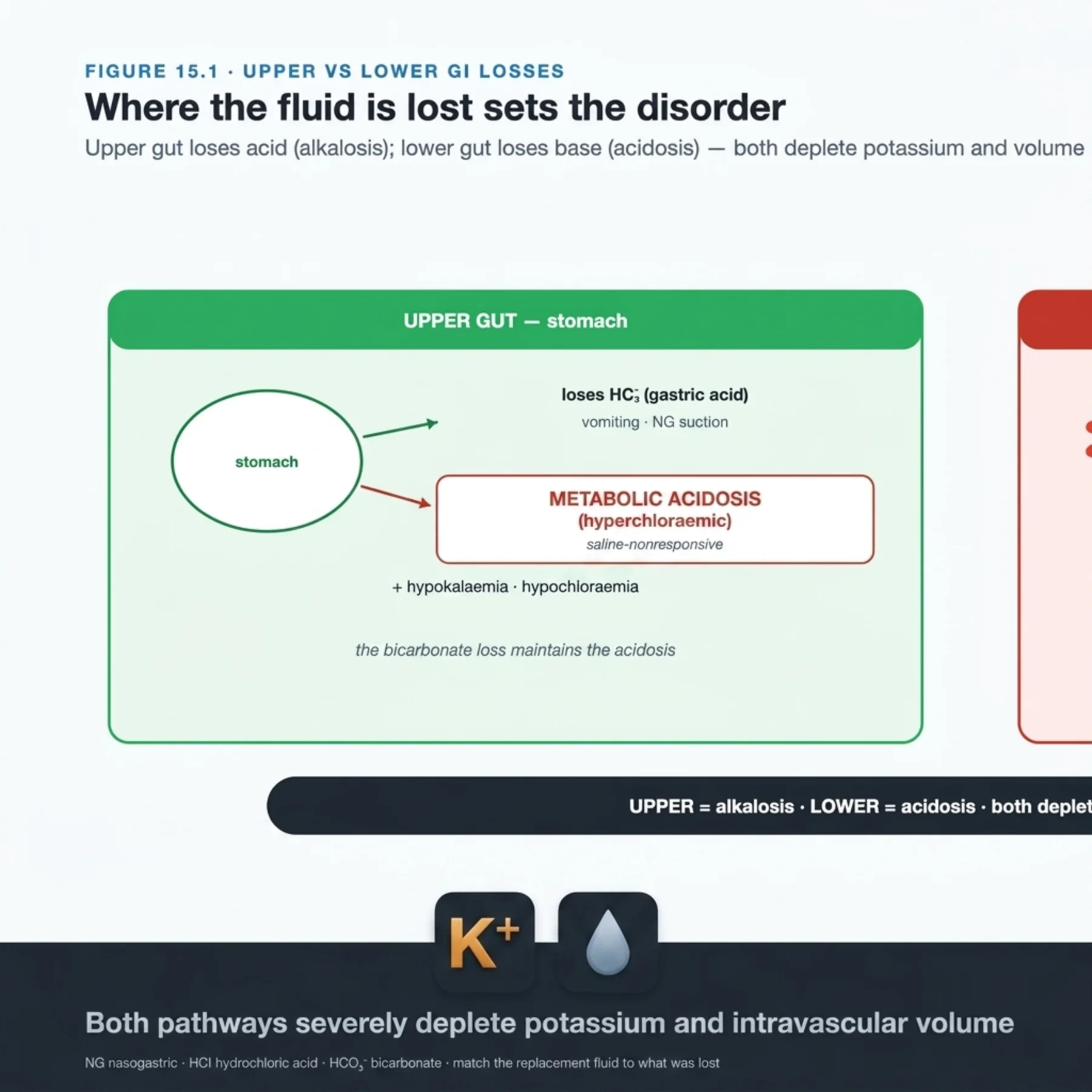
Gastrointestinal losses produce opposite acid-base disturbances depending on where in the gut they come from, and this contrast is one of the most reliable patterns in medicine. Upper gastrointestinal losses — vomiting, nasogastric suction — lose hydrochloric acid, so the body is left with a relative excess of bicarbonate: a metabolic alkalosis, accompanied by hypokalaemia, hypochloraemia, and volume depletion, and (from the last alkalosis chapter) saline-responsive with a low urine chloride. Lower gastrointestinal losses — diarrhoea, intestinal fistulae, a high-output ileostomy — lose bicarbonate-rich fluid, producing a normal-anion-gap metabolic acidosis, again with hypokalaemia and volume depletion (pancreatic and biliary fistulae do the same). So the rule is clean and memorable: upper gut loses acid and causes alkalosis; lower gut loses base and causes acidosis; both deplete potassium and volume. Knowing the source predicts the acid-base disturbance before the gas is drawn, and the replacement is matched to the loss (chloride and potassium for vomiting; bicarbonate and potassium for diarrhoea).
— Endocrine: the adrenal and mineralocorticoid patterns
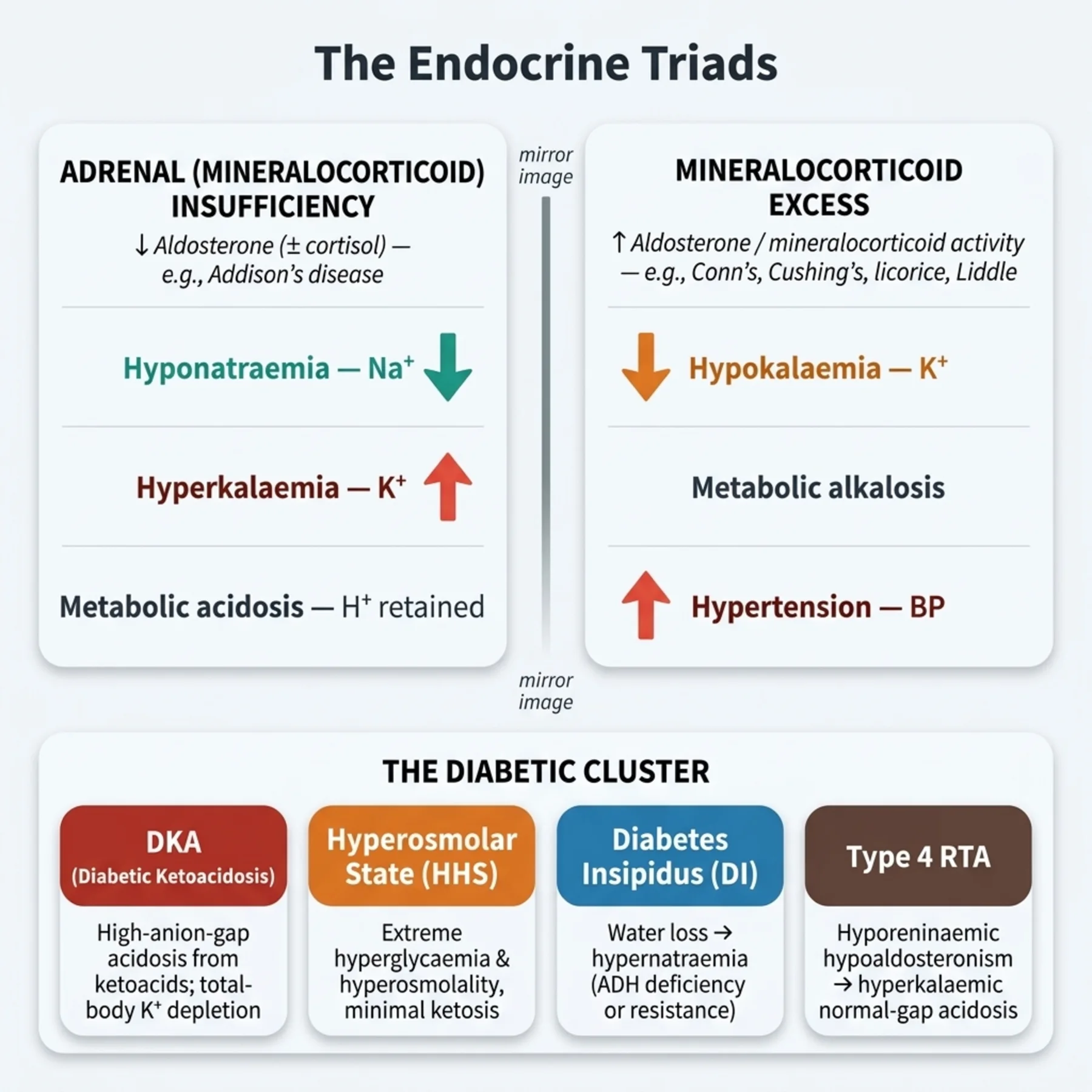
Endocrine disease produces some of the most recognisable disturbance triads. Primary adrenal insufficiency — a deficiency of both aldosterone and cortisol — causes hyponatraemia (loss of cortisol's permissive effect and volume depletion driving ADH), hyperkalaemia (aldosterone deficiency impairing potassium excretion), and a metabolic acidosis (impaired acid excretion) — a triad that, with hypotension and hypoglycaemia, should trigger the diagnosis and treatment with glucocorticoid (and fluids). Its mirror, mineralocorticoid excess (Conn's syndrome and others), causes hypokalaemia, metabolic alkalosis, and hypertension — the saline-resistant alkalosis pattern. The diabetic disturbances are a cluster: diabetic ketoacidosis (a high-gap acidosis with the characteristic total-body potassium depletion despite a high serum level), the hyperosmolar hyperglycaemic state (profound hyperglycaemia and hypernatraemia), diabetes insipidus (hypernatraemia), and the type 4 renal tubular acidosis of hyporeninaemic hypoaldosteronism (a normal-gap acidosis with hyperkalaemia). Each is recognisable, and each is treated by its endocrine cause alongside the disturbance.
— Refeeding syndrome: the insulin surge
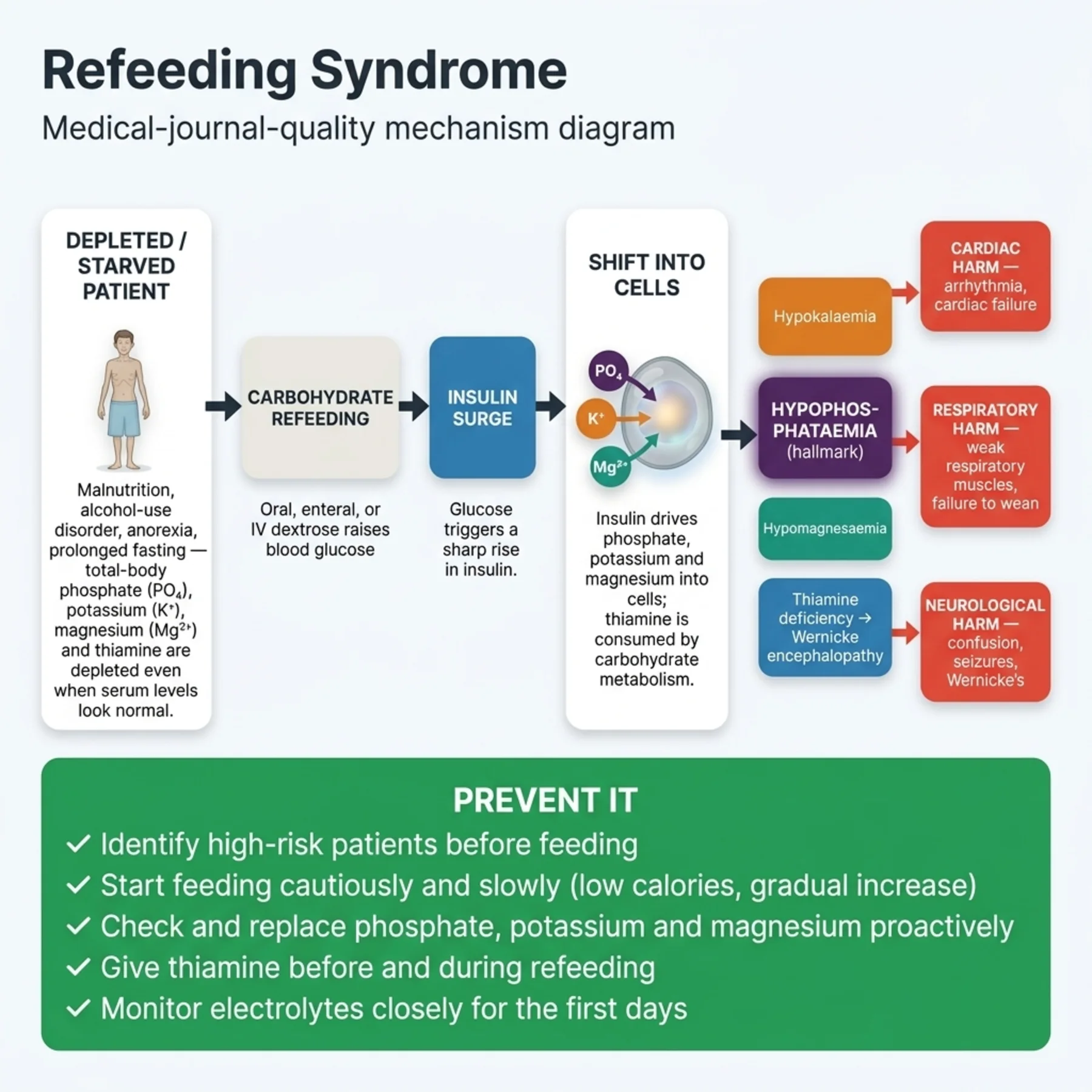
Refeeding syndrome is the disturbance of feeding the starved, and it is both predictable and dangerous. When a malnourished or alcoholic patient — whose intracellular phosphate, potassium, and magnesium stores are depleted — is given carbohydrate, the resulting insulin surge drives phosphate, potassium, and magnesium into cells, and thiamine is consumed in carbohydrate metabolism. The hallmark is hypophosphataemia, which can be profound and cause cardiac arrhythmia, respiratory failure, and neurological harm; hypokalaemia and hypomagnesaemia accompany it, and thiamine deficiency can precipitate Wernicke encephalopathy. Because the mechanism is predictable, the syndrome is preventable: identify the high-risk patient (severe malnutrition, alcoholism, prolonged starvation), start feeding cautiously at a low caloric rate, monitor and replace phosphate, potassium, and magnesium, and give thiamine before or with the feeding. The error is to feed a starved patient fully without anticipation — the very treatment that should help becomes the harm.
— The setting as a diagnostic shortcut
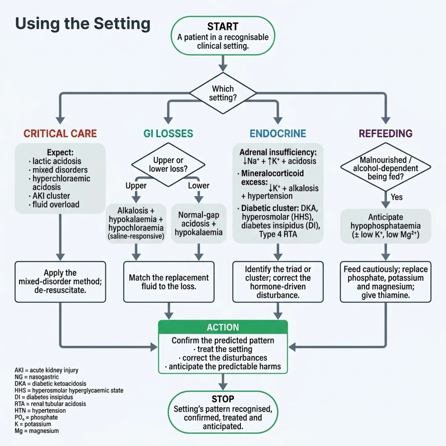
The unifying method of the chapter is to let the setting predict the pattern. Faced with a vomiting patient, expect and look for the alkalosis-hypokalaemia-hypochloraemia signature; with diarrhoea, the acidosis-hypokalaemia pattern; with the hypotensive, hyperkalaemic, hyponatraemic patient, suspect adrenal insufficiency; with the refed malnourished patient, watch the phosphate. This is not a substitute for the systematic methods — the acid-base steps, the electrolyte work-ups — but a powerful accelerant: the setting raises the prior probability of a specific pattern, the systematic method confirms it, and the cause-directed treatment follows. The shortcut also primes anticipation, which is where it saves lives: the refeeding hypophosphataemia and the critical-care overload are not diagnosed after they cause harm but anticipated and prevented because the setting predicted them. Recognising the setting, predicting the pattern, confirming it, and anticipating the predictable harms — that is the discipline.
— Treating across the settings
Treatment in every setting rests on the same two-part principle: treat the underlying setting or cause, and correct the specific disturbances by their own rules (the chapters on each). In critical care, that means restoring perfusion, managing the acute kidney injury, and de-resuscitating the overload. In gastrointestinal losses, it means stemming the loss and replacing the matched fluid and electrolytes — chloride and potassium for upper losses, bicarbonate and potassium for lower. In endocrine disease, it means the hormone (glucocorticoid for adrenal insufficiency, insulin for ketoacidosis) plus the disturbance correction. In refeeding, it means cautious feeding with anticipatory replacement and thiamine. The third element — anticipation — is what distinguishes expert management in these settings: the predictable disturbances (refeeding hypophosphataemia, critical-care overload, the potassium fall in treated ketoacidosis) are pre-empted rather than chased. The setting predicts; the clinician anticipates; the harm is prevented.
| 04 | PHASE A · LEVEL 4 · ORIENTATION & KNOWLEDGE Reference Tables |
Table 15.1 — Critical-care patterns
| Pattern | Detail |
| Lactic acidosis | Hypoperfusion/shock — high-gap acidosis, often mixed |
| Hyperchloraemic acidosis | Large-volume saline resuscitation |
| AKI cluster | Metabolic acidosis, hyperkalaemia, hyperphosphataemia |
| Fluid overload | Resuscitation → de-resuscitation; overload is harmful |
Table 15.2 — Gastrointestinal losses: upper vs lower
| Upper (vomiting, NG) | Lower (diarrhoea, fistula, ileostomy) | |
| What is lost | Hydrochloric acid | Bicarbonate-rich fluid |
| Acid-base | Metabolic ALKALOSIS | Metabolic ACIDOSIS (normal gap) |
| Also | Hypokalaemia, hypochloraemia, volume depletion | Hypokalaemia, volume depletion |
Table 15.3 — Endocrine patterns
| Disorder | Pattern |
| Adrenal insufficiency | Hyponatraemia + HYPERkalaemia + metabolic acidosis |
| Mineralocorticoid excess | Hypokalaemia + metabolic alkalosis + hypertension |
| Diabetic ketoacidosis | High-gap acidosis + total-body K depletion |
| Hyperosmolar state / DI | Hyperglycaemia, hypernatraemia / hypernatraemia |
| Type 4 RTA | Normal-gap acidosis + hyperkalaemia (hyporeninaemic hypoaldosteronism) |
Table 15.4 — Refeeding syndrome
| Element | Detail |
| Trigger | Feeding the malnourished/alcoholic — insulin surge |
| Hallmark | Hypophosphataemia (± hypokalaemia, hypomagnesaemia) |
| Plus | Thiamine deficiency → Wernicke; cardiac/respiratory/neuro harm |
| Prevention | Identify high-risk; feed cautiously; replace PO4/K/Mg; give thiamine |
Table 15.5 — The setting predicts the pattern
| Setting | Expect |
| Vomiting / NG suction | Alkalosis + hypokalaemia + hypochloraemia |
| Diarrhoea / ileostomy | Normal-gap acidosis + hypokalaemia |
| Hypotension + hyperkalaemia + hyponatraemia | Adrenal insufficiency |
| Refed malnourished patient | Hypophosphataemia |
Table 15.6 — Treatment principles
| Principle | Detail |
| Treat the setting | Perfusion, hormone, stem the loss, cautious feeding |
| Correct the disturbance | By its own rules (the relevant chapters) |
| Match the replacement | Chloride/K (upper GI); bicarbonate/K (lower GI) |
| Anticipate | Refeeding hypophosphataemia; critical-care overload; K fall in treated DKA |
| 05 | PHASE B · LEVEL 5 · VISUALISE & MAP Imaging & Flowchart Specifications |
| 06 | PHASE B · LEVEL 6 · VISUALISE & MAP Concept Maps |
Each chain runs from physiology to a named bedside action; read the arrows as “leads to.”
Upper vs lower gut. Upper gut loses HCl → metabolic alkalosis; lower gut loses bicarbonate → normal-gap acidosis; both lose potassium and volume → ACTION: predict the acid-base disturbance from the source and match the replacement.
Adrenal insufficiency. Aldosterone + cortisol deficiency → hyponatraemia + hyperkalaemia + acidosis (+ hypotension, hypoglycaemia) → ACTION: recognise the triad and treat with glucocorticoid and fluids.
Refeeding. Starvation depletes intracellular phosphate/potassium/magnesium → carbohydrate refeeding → insulin surge → those shift into cells → hypophosphataemia → ACTION: anticipate, feed cautiously, replace, and give thiamine.
Critical-care mixtures. Hypoperfusion (lactic acidosis) + saline (hyperchloraemic) + AKI + ventilation changes → mixed disorders + overload → ACTION: apply the mixed-disorder method and de-resuscitate.
The setting predicts. Each setting's pathophysiology dictates a characteristic pattern → the setting raises the prior probability → ACTION: predict, confirm with the systematic method, and anticipate the predictable harms.
| 07 | PHASE B · LEVEL 7 · VISUALISE & MAP Decision Pathways |
| R1 | IF a patient is in a recognisable setting, THEN predict the characteristic disturbance pattern, then confirm it with the systematic method. |
| R2 | IF the losses are upper gastrointestinal (vomiting, NG), THEN expect a metabolic alkalosis with hypokalaemia and hypochloraemia, and replace chloride and potassium. |
| R3 | IF the losses are lower gastrointestinal (diarrhoea, fistula, ileostomy), THEN expect a normal-gap acidosis with hypokalaemia, and replace bicarbonate and potassium. |
| R4 | IF a patient is hyponatraemic, hyperkalaemic, and acidotic (with hypotension), THEN suspect adrenal insufficiency and treat with glucocorticoid and fluids. |
| R5 | IF a patient is hypokalaemic and alkalotic with hypertension, THEN suspect mineralocorticoid excess. |
| R6 | IF feeding a malnourished or alcoholic patient, THEN anticipate refeeding syndrome — feed cautiously, replace phosphate/potassium/magnesium, and give thiamine. |
| R7 | IF managing a critically ill patient, THEN expect mixed disorders and overload — apply the mixed-disorder method and de-resuscitate when stable. |
| R8 | IF in any setting, THEN treat the underlying setting, correct the disturbances by their rules, and anticipate the predictable ones. |
| 08 | PHASE C · LEVEL 8 · CLINICAL REASONING Clinical Cases |
| CASE 1 | UP OR DOWN? The gut predicts the disturbance Upper versus lower GI losses |
Presentation
Two patients present with gastrointestinal losses: one with persistent vomiting, the other with a high-output ileostomy. Both are hypokalaemic and volume-depleted, and the team expects the same acid-base disturbance in each.
❖ Pause and reflect Will both have the same acid-base disturbance? |
Analysis
No — they will be opposite. The vomiting patient loses hydrochloric acid, producing a metabolic alkalosis (with hypokalaemia, hypochloraemia, volume depletion, and a low urine chloride — saline-responsive). The ileostomy patient loses bicarbonate-rich intestinal fluid, producing a normal-anion-gap metabolic acidosis (with hypokalaemia and volume depletion). The source of the loss predicts the acid-base disturbance — upper gut alkalosis, lower gut acidosis — even though both share the hypokalaemia and volume depletion. The replacements differ accordingly.
Plan
For the vomiting patient, give saline with potassium chloride (correcting the alkalosis, chloride, and potassium); for the ileostomy patient, replace volume, bicarbonate, and potassium (correcting the acidosis). Match the replacement to the source and its acid-base disturbance.
Teaching point
Upper gastrointestinal losses cause alkalosis, lower cause acidosis — both cause hypokalaemia and volume depletion; the source predicts the disturbance.
Cross-reference
Exercises rules R2 and R3; the upper-versus-lower concept map; Figure 15.1; Tables 15.2, 15.6; alkalosis in Chapter 12, acidosis in Chapter 11.
| CASE 2 | THE TELLTALE TRIAD Recognise the pattern Adrenal insufficiency |
Presentation
A hypotensive patient is found to have hyponatraemia, hyperkalaemia, and a mild metabolic acidosis, with a history of fatigue and weight loss. The team is treating the electrolytes individually.
❖ Pause and reflect What single diagnosis ties these disturbances together? |
Analysis
The triad of hyponatraemia, hyperkalaemia, and metabolic acidosis with hypotension points to adrenal insufficiency. Aldosterone deficiency impairs potassium and acid excretion (hyperkalaemia, acidosis) and causes volume depletion, while cortisol deficiency contributes to the hyponatraemia (and the hypotension and hypoglycaemia). Treating each electrolyte in isolation misses the unifying diagnosis and the life-saving treatment — glucocorticoid replacement — which corrects the whole pattern. The setting and the triad are the diagnostic shortcut.
Plan
Recognise the adrenal insufficiency, confirm it, and treat with glucocorticoid (hydrocortisone) and fluids, which correct the pattern; address the hyperkalaemia acutely if dangerous. Treat the unifying cause, not just the individual electrolytes.
Teaching point
Hyponatraemia + hyperkalaemia + acidosis with hypotension is adrenal insufficiency — treat the cause (glucocorticoid), not just the electrolytes.
Cross-reference
Exercises rule R4; the adrenal-insufficiency concept map; Figure 15.2; Table 15.3.
| CASE 3 | FEEDING THE STARVED Anticipate the phosphate Refeeding syndrome |
Presentation
A severely malnourished patient with alcohol-use disorder is admitted, and the team plans to start full nutritional support immediately to correct the malnutrition.
❖ Pause and reflect What will happen if this patient is fed fully without precaution? |
Analysis
Full feeding without precaution risks refeeding syndrome. The patient's intracellular phosphate, potassium, and magnesium are depleted; carbohydrate triggers an insulin surge that drives these into cells, causing a profound hypophosphataemia (the hallmark) with hypokalaemia and hypomagnesaemia, and thiamine is consumed — risking cardiac arrhythmia, respiratory failure, and Wernicke encephalopathy. This is predictable and preventable: the high-risk patient should be fed cautiously at a low caloric rate, with monitoring and replacement of phosphate, potassium, and magnesium, and thiamine given before or with the feeding.
Plan
Start feeding cautiously at a low caloric rate, monitor and replace phosphate, potassium, and magnesium, and give thiamine before or with the feeding; escalate calories slowly. Anticipate refeeding syndrome in every high-risk patient.
Teaching point
Anticipate refeeding syndrome in the malnourished/alcoholic — hypophosphataemia is the hallmark; feed cautiously, replace, and give thiamine.
Cross-reference
Exercises rule R6; the refeeding concept map; Figure 15.3; Table 15.4; hypophosphataemia in Chapter 9.
| CASE 4 | THE ICU MIXTURE Many disorders at once Critical-care patterns |
Presentation
A septic, resuscitated ICU patient has a lactic acidosis, a hyperchloraemic component from large-volume saline, an evolving acute kidney injury, and is now becoming oedematous and overloaded.
❖ Pause and reflect How do you make sense of this tangle of disturbances? |
Analysis
This is the typical critical-care mixture, and it is parsed with the systematic and mixed-disorder methods. There is a high-gap lactic acidosis (hypoperfusion), a superimposed hyperchloraemic normal-gap acidosis (the saline), the acid-base and electrolyte consequences of the AKI, and now fluid overload from the resuscitation. The management is multi-pronged: restore perfusion and treat the sepsis (for the lactic acidosis), prefer balanced fluids (limiting the hyperchloraemic acidosis), manage the AKI, and de-resuscitate the overload now that the patient is stabilising. The critically ill patient rarely has one disorder.
Plan
Apply the mixed-disorder method to name each disturbance, restore perfusion and treat the sepsis, switch to balanced fluids, manage the AKI, and begin de-resuscitation of the overload. Expect and manage multiple coexisting disorders.
Teaching point
The critically ill patient has mixed disorders and overload — apply the mixed-disorder method, prefer balanced fluids, and de-resuscitate.
Cross-reference
Exercises rule R7; the critical-care concept map; Table 15.1; mixed disorders in Chapter 13; fluids in Chapter 2; overload in Volume 5.
| 09 | PHASE C · LEVEL 9 · CLINICAL REASONING Clinical Implications |
One triad per mechanism the narrative exposed: the physiology, why it matters, and the bedside move.
MECHANISM Upper gut loses hydrochloric acid while lower gut loses bicarbonate-rich fluid. |
WHY IT MATTERS The source of gastrointestinal loss determines whether the disturbance is alkalosis or acidosis. |
ACTION Predict the acid-base disturbance from the source and match the replacement. |
MECHANISM Adrenal insufficiency removes both aldosterone and cortisol. |
WHY IT MATTERS This produces the recognisable triad of hyponatraemia, hyperkalaemia, and acidosis. |
ACTION Recognise the triad and treat the cause with glucocorticoid, not just the electrolytes. |
MECHANISM Refeeding drives an insulin surge that shifts phosphate, potassium, and magnesium into cells. |
WHY IT MATTERS The malnourished patient develops a dangerous, predictable hypophosphataemia. |
ACTION Anticipate it — feed cautiously, replace, and give thiamine. |
MECHANISM Critical illness combines hypoperfusion, saline, acute kidney injury, and ventilation changes. |
WHY IT MATTERS The result is mixed disorders and fluid overload, not a single disturbance. |
ACTION Apply the mixed-disorder method and de-resuscitate. |
MECHANISM Each setting's pathophysiology dictates a characteristic disturbance pattern. |
WHY IT MATTERS The setting raises the prior probability of a specific pattern. |
ACTION Predict from the setting, confirm with the method, and anticipate the predictable harms. |
| 10 | PHASE C · LEVEL 10 · CLINICAL REASONING Clinical Pearls |
| The clinical setting predicts the disturbance pattern — a diagnostic shortcut. | Upper GI (vomiting/NG): metabolic ALKALOSIS + hypoK + hypochloraemia. |
| Lower GI (diarrhoea/fistula/ileostomy): normal-gap ACIDOSIS + hypoK. | Both GI: hypokalaemia + volume depletion. |
| Match replacement: Cl/K (upper); bicarbonate/K (lower). | Adrenal insufficiency: hyponatraemia + HYPERkalaemia + acidosis (+ hypotension). |
| Treat adrenal insufficiency with glucocorticoid + fluids. | Mineralocorticoid excess: hypoK + alkalosis + hypertension. |
| DKA: high-gap acidosis + total-body K depletion. | Type 4 RTA: normal-gap acidosis + hyperkalaemia. |
| Refeeding: insulin surge → hypoPHOSPHATAEMIA (hallmark) + hypoK + hypoMg. | Refeeding: + thiamine deficiency (Wernicke); cardiac/respiratory/neuro harm. |
| Prevent refeeding: cautious feeding, replace PO4/K/Mg, give thiamine. | Critical care: lactic acidosis, mixed disorders, hyperchloraemic, AKI, overload. |
| ICU: apply mixed-disorder method; de-resuscitate. | Treat the setting + correct disturbances + ANTICIPATE the predictable. |
| 11 | PHASE D · LEVEL 11 · SAFETY & EVIDENCE Red Flags & Never-Do |
Panel A — Red flags
| ▲ | Hyponatraemia + hyperkalaemia + acidosis with hypotension — adrenal insufficiency; give glucocorticoid. |
| ▲ | A malnourished/alcoholic patient about to be fed — refeeding risk; anticipate hypophosphataemia and give thiamine. |
| ▲ | A vomiting patient with alkalosis — saline-responsive; replace chloride and potassium. |
| ▲ | A high-output ileostomy or diarrhoea with acidosis — replace bicarbonate and potassium. |
| ▲ | A resuscitated ICU patient becoming oedematous — fluid overload; begin de-resuscitation. |
Panel B — Never do
| ✖ NEVER — treat the electrolytes of adrenal insufficiency without giving glucocorticoid. |
| ✖ NEVER — feed a high-risk malnourished patient fully without anticipating refeeding syndrome. |
| ✖ NEVER — assume gastrointestinal losses cause the same acid-base disturbance regardless of source. |
| ✖ NEVER — leave critical-care overload unaddressed once the patient stabilises. |
| 12 | PHASE D · LEVEL 12 · SAFETY & EVIDENCE Common Pitfalls |
Pitfall 1 — Same disturbance for all GI losses
| ✖ | WRONG Expecting the same acid-base disturbance from vomiting and diarrhoea. |
| ✓ | RIGHT Predicting alkalosis from upper losses and acidosis from lower. |
| ✉ | WHY Upper gut loses acid, lower gut loses base. |
Pitfall 2 — Missing adrenal insufficiency
| ✖ | WRONG Treating the hyponatraemia, hyperkalaemia, and acidosis separately. |
| ✓ | RIGHT Recognising the triad as adrenal insufficiency and giving glucocorticoid. |
| ✉ | WHY A single hormone deficiency explains the whole pattern. |
Pitfall 3 — Feeding without anticipation
| ✖ | WRONG Starting full feeding in a malnourished patient. |
| ✓ | RIGHT Feeding cautiously with anticipatory replacement and thiamine. |
| ✉ | WHY The insulin surge causes a dangerous, predictable hypophosphataemia. |
Pitfall 4 — Treating the ICU patient as having one disorder
| ✖ | WRONG Naming a single disturbance in a critically ill patient. |
| ✓ | RIGHT Applying the mixed-disorder method to name them all. |
| ✉ | WHY Critical illness produces multiple coexisting disturbances. |
Pitfall 5 — Ignoring overload
| ✖ | WRONG Continuing resuscitation fluids after stabilisation. |
| ✓ | RIGHT De-resuscitating once the patient is stable. |
| ✉ | WHY Fluid overload is harmful and must be actively removed. |
| 13 | PHASE D · LEVEL 13 · SAFETY & EVIDENCE Evidence Grading |
GRADE A HIGH CONFIDENCE The effect is real and the estimate is stable. RCTs at low risk of bias; multiple concordant prospective cohorts; meta-analyses. |
GRADE B MODERATE CONFIDENCE The effect is likely real but may shift with new data. Observational studies, registries, mechanistic human studies. |
GRADE C LOW CONFIDENCE Rests on physiology, reasoning, or consensus rather than outcomes. Pathophysiological reasoning; extrapolation; consensus without outcomes. |
Graded statements (by evidence type)
| Statement | Grade | Basis (evidence type) |
| Upper GI losses cause alkalosis; lower GI losses cause acidosis. | A | Established physiology |
| Adrenal insufficiency causes hyponatraemia, hyperkalaemia, and acidosis. | A | Established physiology |
| Refeeding causes insulin-mediated hypophosphataemia. | A | Established physiology and clinical data |
| Refeeding syndrome is preventable with cautious feeding and replacement. | A | Clinical guidelines |
| Critical illness commonly produces mixed acid-base disorders. | A | Clinical observation |
| Fluid overload in the critically ill is associated with worse outcomes. | B | Observational and trial data |
| The clinical setting predicts the disturbance pattern. | A | Established physiology |
| 14 | PHASE E · LEVEL 14 · PATIENT DECISIONS Absolute Risk in Natural Frequency |
Natural-frequency estimates for orientation, from these settings; they vary with severity and risk. They convey the size of the decisions, expressed per 100 comparable patients.
| Per 100 patients… | Outcome | Roughly how many | See |
| High-risk patients fed without precaution | Develop refeeding syndrome | A meaningful share — hence anticipate | L13 row 3 |
| Refeeding-risk patients managed with a protocol | Avoid severe hypophosphataemia | Most | L13 row 4 |
| Adrenal insufficiency given glucocorticoid | Have the whole pattern corrected | Most | L13 row 2 |
| Critically ill left fluid-overloaded | Have a worse outcome | More than the de-resuscitated | L13 row 6 |
★ How to read these Read these as orientation, not promises; outcomes depend on severity and risk. The stable signals: refeeding is preventable with a protocol, glucocorticoid corrects adrenal insufficiency, and unaddressed overload harms. Communicate them as people out of 100, not as a hazard ratio. |
| 17 | PHASE F · LEVEL 17 · APPLY & TEST Documentation Templates |
Paste-ready notes. Tick the boxes that apply and delete the rest; make the predicted pattern, the matched replacement, and the anticipation explicit.
Template 1 — Gastrointestinal-loss assessment and replacement
Template 2 — Refeeding risk and protocol
| 18 | PHASE F · LEVEL 18 · APPLY & TEST Cheat Sheet |
| The setting predicts the pattern — a diagnostic shortcut. | Upper GI (vomiting/NG): alkalosis + hypoK + hypochloraemia. |
| Lower GI (diarrhoea/fistula): normal-gap acidosis + hypoK. | Both GI: hypokalaemia + volume depletion. |
| Match: Cl/K (upper); bicarbonate/K (lower). | Adrenal insufficiency: hypoNa + hyperK + acidosis + hypotension. |
| Treat adrenal insufficiency: glucocorticoid + fluids. | Mineralocorticoid excess: hypoK + alkalosis + HTN. |
| DKA: high-gap acidosis + total-body K depletion. | Type 4 RTA: normal-gap acidosis + hyperK. |
| Refeeding: insulin → hypophosphataemia (hallmark) + hypoK + hypoMg. | Refeeding: + thiamine deficiency (Wernicke). |
| Prevent refeeding: cautious feed, replace PO4/K/Mg, thiamine. | Critical care: lactic + mixed + hyperchloraemic + AKI + overload. |
| ICU: mixed-disorder method; de-resuscitate. | Treat setting + correct + ANTICIPATE. |
| 19 | PHASE F · LEVEL 19 · APPLY & TEST Flashcards |
| CARD 1 | Q. Why do specific clinical settings produce characteristic disturbance patterns? A. Because the underlying pathophysiology of each setting dictates a predictable combination of fluid, electrolyte, and acid-base disturbances — making the setting a powerful diagnostic shortcut. DETAILED. Predict the pattern, then confirm and treat. CLINICAL. Use the setting to raise the prior probability of a specific pattern. |
| CARD 2 | Q. How do upper and lower gastrointestinal losses differ? A. Upper losses (vomiting, nasogastric suction) lose hydrochloric acid, causing a metabolic alkalosis with hypokalaemia and hypochloraemia; lower losses (diarrhoea, fistula, ileostomy) lose bicarbonate-rich fluid, causing a normal-gap acidosis with hypokalaemia. Both deplete volume. DETAILED. Upper gut loses acid, lower gut loses base. CLINICAL. Predict the disturbance from the source and match the replacement. |
| CARD 3 | Q. What is the triad of adrenal insufficiency? A. Hyponatraemia, hyperkalaemia, and a metabolic acidosis (with hypotension and hypoglycaemia), from combined aldosterone and cortisol deficiency. DETAILED. A single hormone deficiency explains the whole pattern. CLINICAL. Recognise the triad and treat with glucocorticoid and fluids. |
| CARD 4 | Q. What is the pattern of mineralocorticoid excess? A. Hypokalaemia, a metabolic alkalosis, and hypertension — the mirror of adrenal insufficiency. DETAILED. It is the saline-resistant alkalosis pattern. CLINICAL. Recognise it and investigate with renin and aldosterone. |
| CARD 5 | Q. What causes refeeding syndrome and what is its hallmark? A. Feeding a malnourished or alcoholic patient triggers an insulin surge that shifts phosphate, potassium, and magnesium into cells, with thiamine consumed; the hallmark is hypophosphataemia, risking cardiac, respiratory, and neurological harm. DETAILED. It is predictable and preventable. CLINICAL. Anticipate it — feed cautiously, replace, and give thiamine. |
| CARD 6 | Q. How is refeeding syndrome prevented? A. By identifying high-risk patients, starting feeding cautiously at a low caloric rate, monitoring and replacing phosphate, potassium, and magnesium, and giving thiamine before or with the feeding. DETAILED. The error is full feeding without anticipation. CLINICAL. Use a refeeding protocol in every high-risk patient. |
| CARD 7 | Q. What are the characteristic critical-care disturbances? A. Lactic acidosis from hypoperfusion (often as part of a mixed disorder), hyperchloraemic acidosis from saline, the acid-base and electrolyte cluster of acute kidney injury, and fluid overload. DETAILED. They are usually mixed, not single. CLINICAL. Apply the mixed-disorder method and de-resuscitate the overload. |
| CARD 8 | Q. What is the unifying treatment principle across these settings? A. Treat the underlying setting or cause, correct the specific disturbances by their own rules, and anticipate the predictable disturbances (refeeding hypophosphataemia, critical-care overload, the potassium fall in treated ketoacidosis). DETAILED. Anticipation distinguishes expert management. CLINICAL. Treat the setting, correct the disturbance, and anticipate. |
| 20 | PHASE F · LEVEL 20 · APPLY & TEST One-Minute Preceptor |
| SCENE 1 | The intern expecting one GI pattern |
GET A COMMITMENT. “You expect the same acid-base disturbance in this vomiting patient and that ileostomy patient — why?”
PROBE FOR EVIDENCE. “They're both losing GI fluid” — ask: “What does the stomach lose, and what does the intestine lose?”
TEACH A GENERAL RULE. Upper gut loses acid (alkalosis), lower gut loses bicarbonate (acidosis) — opposite disturbances, though both deplete potassium and volume.
REINFORCE WHAT WAS RIGHT. Recognising both as GI losses was correct.
CORRECT A MISTAKE. Predict alkalosis for the vomiting, acidosis for the ileostomy, and match the replacement.
| SCENE 2 | The resident about to feed the starved |
GET A COMMITMENT. “You're starting full feeds in this malnourished alcoholic — any concern?”
PROBE FOR EVIDENCE. “He needs the nutrition” — ask: “What does carbohydrate do to insulin, and where does the phosphate go?”
TEACH A GENERAL RULE. Refeeding triggers an insulin surge that shifts phosphate into cells, causing a dangerous hypophosphataemia — feed cautiously, replace, and give thiamine.
REINFORCE WHAT WAS RIGHT. Wanting to correct the malnutrition was right.
CORRECT A MISTAKE. Feed slowly with anticipatory replacement and thiamine.
| 22 | PHASE F · LEVEL 22 · APPLY & TEST Board-Style Questions |
| Q 01 | Why is the clinical setting a useful diagnostic tool in fluid and electrolyte disorders? |
| A | It is not useful |
| B | Each setting's pathophysiology predicts a characteristic disturbance pattern |
| C | It replaces the systematic method |
| D | It only applies to the ICU |
Rationale The setting raises the prior probability of a specific pattern, a powerful shortcut confirmed by the method (Figure 15.A, rule R1). A, C, and D are incorrect. |
| Q 02 | Vomiting and nasogastric suction characteristically cause: |
| A | A normal-gap acidosis |
| B | A metabolic alkalosis with hypokalaemia and hypochloraemia |
| C | A respiratory acidosis |
| D | No disturbance |
Rationale Upper gastrointestinal losses lose hydrochloric acid, causing a saline-responsive metabolic alkalosis (case 1, Table 15.2, rule R2). A is a lower-GI pattern; C and D are wrong. |
| Q 03 | A high-output ileostomy or diarrhoea characteristically causes: |
| A | A metabolic alkalosis |
| B | A normal-anion-gap metabolic acidosis with hypokalaemia |
| C | A respiratory alkalosis |
| D | Hypernatraemia |
Rationale Lower gastrointestinal losses lose bicarbonate-rich fluid, causing a normal-gap acidosis (case 1, Table 15.2, rule R3). A is an upper-GI pattern; C and D are wrong. |
| Q 04 | A hypotensive patient with hyponatraemia, hyperkalaemia, and a metabolic acidosis most likely has: |
| A | Mineralocorticoid excess |
| B | Adrenal insufficiency |
| C | Diabetic ketoacidosis |
| D | Refeeding syndrome |
Rationale This triad with hypotension reflects combined aldosterone and cortisol deficiency — adrenal insufficiency (case 2, Table 15.3, rule R4). A is the opposite; C and D give different patterns. |
| Q 05 | The hallmark electrolyte disturbance of refeeding syndrome is: |
| A | Hyperphosphataemia |
| B | Hypophosphataemia |
| C | Hypernatraemia |
| D | Hypercalcaemia |
Rationale The insulin surge shifts phosphate into cells, causing the characteristic hypophosphataemia (case 3, Table 15.4, rule R6). A is the opposite; C and D are unrelated. |
| Q 06 | How is refeeding syndrome prevented? |
| A | Full feeding immediately |
| B | Cautious feeding with phosphate/potassium/magnesium replacement and thiamine |
| C | Withholding all nutrition |
| D | High-calorie feeding |
Rationale Cautious feeding with anticipatory replacement and thiamine prevents the syndrome (case 3, Table 15.4, rule R6). A and D precipitate it; C is not the answer. |
| Q 07 | The characteristic acid-base picture of critical illness is: |
| A | A single pure disorder |
| B | Mixed disorders (lactic, hyperchloraemic, AKI) with fluid overload |
| C | Always a metabolic alkalosis |
| D | Always a respiratory alkalosis |
Rationale Critical illness combines several disturbances and overload, requiring the mixed-disorder method (case 4, Table 15.1, rule R7). A, C, and D oversimplify. |
| Q 08 | Mineralocorticoid excess (e.g. Conn's syndrome) characteristically causes: |
| A | Hyperkalaemia and acidosis |
| B | Hypokalaemia, metabolic alkalosis, and hypertension |
| C | Hyponatraemia and hypotension |
| D | Hypophosphataemia |
Rationale Mineralocorticoid excess is the mirror of adrenal insufficiency — hypokalaemia, alkalosis, hypertension (Table 15.3, rule R5). A is adrenal insufficiency; C and D are wrong. |
| Q 09 | The unifying treatment principle across these settings is to: |
| A | Treat only the numbers |
| B | Treat the setting, correct the disturbances, and anticipate the predictable ones |
| C | Ignore the cause |
| D | Always start with dialysis |
Rationale Treating the setting, correcting the disturbances, and anticipating the predictable harms is the principle (Table 15.6, rule R8). A, C, and D are incomplete. |