Chapter Preamble
Signals declared
Sig-D — Diagnostic (primary). Separate redistribution from depletion, localise renal versus extrarenal loss, and read the ECG and the magnesium.
Sig-T — Therapeutic (strong). Replacing potassium safely by the right route and rate, replacing magnesium, and treating the cause.
Sig-M — Mechanistic (strong). Potassium's internal and external balance, renal secretion, and the magnesium dependence that makes hypokalaemia refractory.
Levels populated and omitted
Populated (19): L1–L14, L17–L20, L22. The mechanistic signal fires the concept maps (L6) and triads (L9); the therapeutic signal fires the absolute-risk table (L14) and templates (L17); the diagnostic signal drives the tables, rules, cases, pitfalls, and board items.
L15 / L16 preference-sensitive map and SDM scripts — omitted. No Sig-E; correcting hypokalaemia is effective care.
L21 reflective prompts — omitted. No Sig-E/V; the chapter's tensions (redistribution versus depletion, the magnesium trap) are worked through the cases and pitfalls.
| 01 | PHASE A · LEVEL 1 · ORIENTATION & KNOWLEDGE Learning Objectives |
By the end of this chapter you should be able to:
Explain potassium's internal (shift) and external (total-body) balance.
Distinguish redistributive hypokalaemia from true depletion.
Use the urine potassium to localise depletion as renal or extrarenal.
Classify renal potassium loss using acid-base status and blood pressure.
Explain why hypomagnesaemia causes refractory hypokalaemia.
Recognise the ECG changes and the arrhythmia risk, including with digoxin.
Replace potassium safely by the appropriate route and rate, and replace magnesium.
Avoid over-replacement in redistributive hypokalaemia.
| 02 | PHASE A · LEVEL 2 · ORIENTATION & KNOWLEDGE Executive Summary |
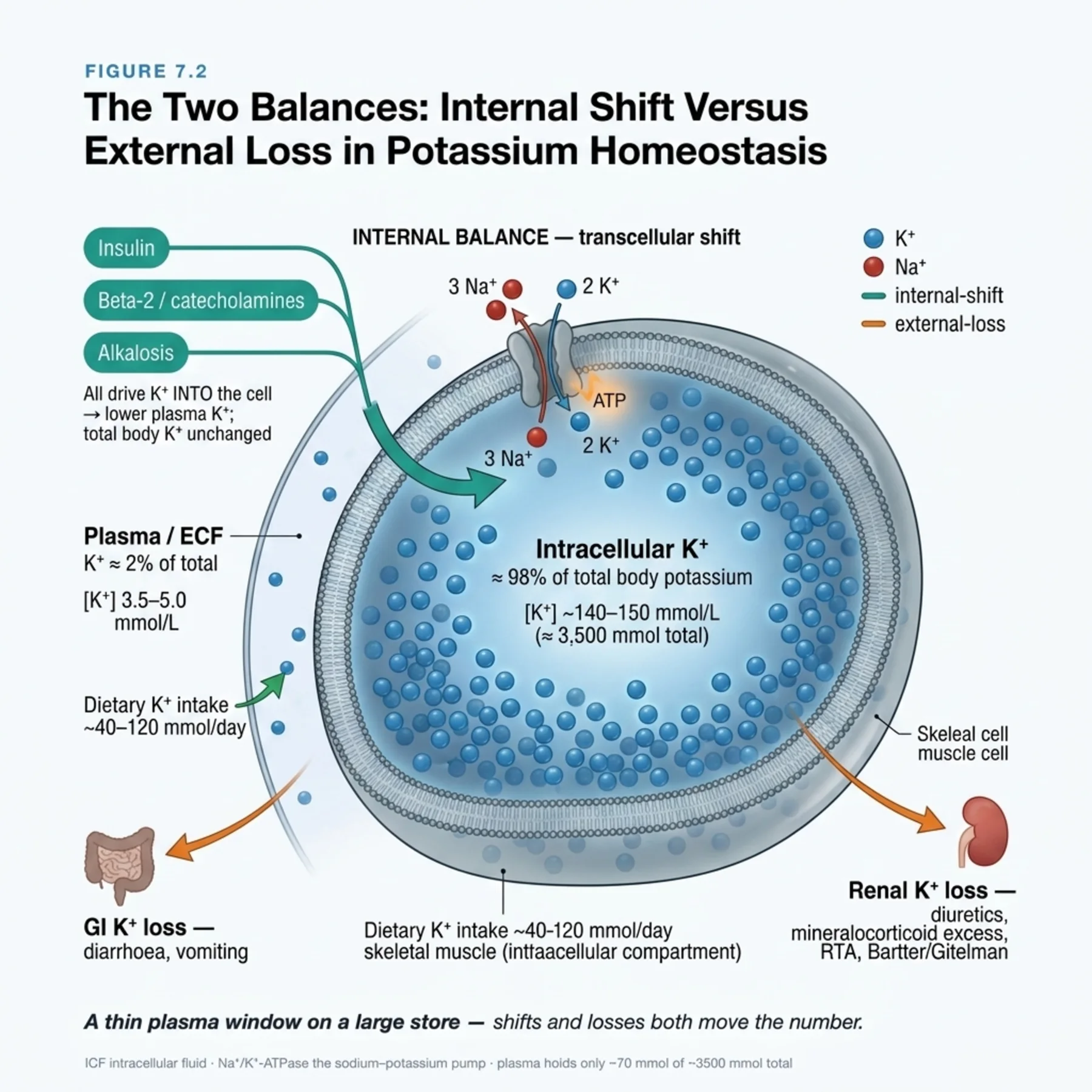
Potassium is overwhelmingly intracellular, so the plasma level reflects only a tiny fraction of total body potassium and small shifts move it a lot.
Internal balance shifts potassium between cells and plasma via the sodium-potassium pump; insulin, beta-adrenergic stimulation, and alkalosis drive it into cells.
External balance is total-body potassium, governed by renal excretion (the main route) and gastrointestinal loss.
Hypokalaemia is therefore either redistribution (a shift into cells) or true depletion (loss exceeding intake).
The first diagnostic step is to separate these, because redistribution corrects when the stimulus resolves and over-replacement then causes rebound hyperkalaemia.
In depletion, the urine potassium localises the loss: low with extrarenal (gastrointestinal) loss or low intake, high with renal loss.
Renal loss is classified by acid-base status and blood pressure — alkalosis with normal or low blood pressure (diuretics, vomiting, Bartter and Gitelman), alkalosis with hypertension (mineralocorticoid excess), and acidosis (renal tubular acidosis).
Hypomagnesaemia causes renal potassium wasting and makes hypokalaemia refractory — the magnesium must be replaced or the potassium will not correct.
The ECG shows U waves, T-wave flattening, ST depression, and QT-QU prolongation, with a risk of arrhythmia that is amplified by digoxin.
Treatment urgency is set by the severity, symptoms, ECG changes, and arrhythmia risk.
Oral potassium is preferred for mild to moderate depletion; intravenous potassium is for severe, symptomatic, or non-oral cases, given at controlled rates and never as a bolus.
Magnesium is replaced whenever it is low, and the underlying cause is treated.
In redistributive hypokalaemia the cause is treated and replacement is cautious, because the potassium will return to the plasma.
Hypokalaemia and digoxin together are a dangerous combination, demanding careful correction.
| 03 | PHASE A · LEVEL 3 · ORIENTATION & KNOWLEDGE Main Narrative |
Potassium lives inside cells. Only about two percent of the body's potassium is in the plasma, so the number on the report is a thin window onto a vast intracellular store, and a small shift across the cell membrane — or a modest total deficit — moves it dramatically. That single fact organises hypokalaemia: a low potassium is either a shift into cells or a true loss of body potassium, and almost everything in diagnosis and treatment flows from telling those two apart and respecting the magnesium that quietly governs the kidney's handling of potassium.
— Two balances: internal and external
Potassium homeostasis runs on two systems. Internal balance is the rapid shift of potassium between the cells and the extracellular fluid, mediated by the sodium-potassium-ATPase pump. Insulin and beta-adrenergic stimulation activate the pump and drive potassium into cells, and alkalosis does the same as cells take up potassium in exchange for hydrogen ions; acidosis and cell breakdown move potassium out. This internal system buffers the plasma minute to minute but does not change total body potassium — it just redistributes it. External balance is the total-body content, set by the difference between intake and output, with the kidney as the dominant route of excretion (potassium is secreted in the distal nephron under the influence of aldosterone, distal sodium delivery and flow, and the lumen-negative voltage generated by sodium reabsorption) and the gut as a secondary route. Hypokalaemia is a disorder of one or both systems: a redistributive shift into cells, a true depletion from loss exceeding intake, or both.
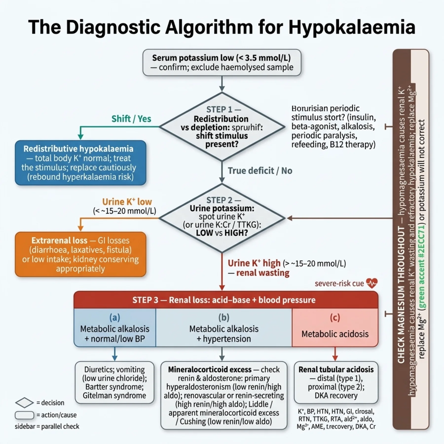
— Step one: redistribution or depletion?
The first and most consequential question is whether the low potassium is a shift or a true deficit, because the two are managed quite differently. Redistributive hypokalaemia — from insulin (as in the treatment of diabetic ketoacidosis), beta-agonists, alkalosis, the hypokalaemic and thyrotoxic periodic paralyses, refeeding, and states of high cellular uptake — reflects potassium hiding inside cells, not lost from the body, so when the stimulus resolves the potassium returns to the plasma. Treating redistribution as if it were depletion, by aggressive replacement, sets up rebound hyperkalaemia once the shift reverses — a real danger in periodic paralysis, where the total body potassium is normal. True depletion, by contrast, is a genuine loss that must be replaced. The clinical context usually distinguishes them (a clear shift stimulus versus a source of loss), and recognising redistribution prevents the over-replacement error.
— Localising depletion: the urine potassium
Once true depletion is established, the next question is where the potassium is being lost, answered by the urine potassium. When the kidney is appropriately conserving potassium, the urine potassium is low (a 24-hour value under about 15 to 20 mmol, or a low spot potassium-to-creatinine ratio), pointing to extrarenal loss — gastrointestinal, usually diarrhoea, since lower gastrointestinal fluid is potassium-rich — or to poor intake. When the urine potassium is high despite hypokalaemia, the kidney is inappropriately wasting potassium, and the loss is renal. This single measurement splits the depletion causes into the extrarenal group (treated by addressing the loss and replacing potassium) and the much larger and more varied renal group, which then needs further classification.
— Classifying renal loss: acid-base and blood pressure
Renal potassium wasting is classified using two readily available pieces of information: the acid-base status and the blood pressure. Renal loss with a metabolic alkalosis and a normal or low blood pressure points to diuretics, to vomiting (which causes renal potassium loss through volume depletion and secondary hyperaldosteronism rather than direct gastric loss), and to the inherited tubulopathies Bartter and Gitelman syndromes. Renal loss with a metabolic alkalosis and hypertension points to mineralocorticoid excess — primary hyperaldosteronism (Conn's syndrome), Cushing's syndrome, Liddle syndrome, and renovascular disease — investigated with the renin and aldosterone. Renal loss with a metabolic acidosis points to the renal tubular acidoses (distal type 1 and proximal type 2) and to states such as treated diabetic ketoacidosis and ureteral diversion. This two-axis classification — acid-base and blood pressure — turns the long list of renal causes into a short, navigable scheme that the acid-base chapters will reinforce.
— The magnesium trap
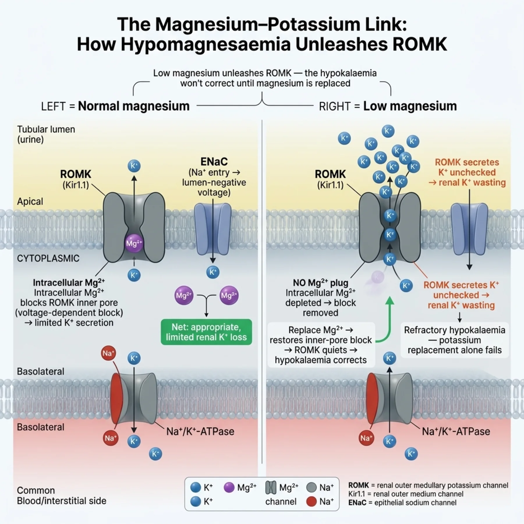
One mechanism deserves singling out because it defeats treatment when missed: hypomagnesaemia causes refractory hypokalaemia. Magnesium normally inhibits the ROMK potassium channel in the distal nephron; when magnesium is depleted, that inhibition is lifted, the channel secretes potassium unchecked, and the kidney wastes potassium. The clinical consequence is that a hypomagnesaemic patient's hypokalaemia simply will not correct with potassium replacement alone — the kidney keeps excreting what is given — until the magnesium is also replaced. Because hypomagnesaemia and hypokalaemia share causes (diuretics, diarrhoea, alcohol, certain drugs), they frequently coexist, and checking and replacing magnesium is a near-universal step in persistent hypokalaemia. Forgetting it is the commonest reason hypokalaemia 'refuses' to respond.
— The ECG and the danger
Hypokalaemia is not merely a number; it changes cardiac membrane excitability and threatens arrhythmia. The electrocardiogram shows a recognisable sequence — flattening then inversion of the T wave, ST-segment depression, prominent U waves, and prolongation of the QT (more precisely the QU) interval — and severe hypokalaemia predisposes to ventricular arrhythmias. The danger is sharply amplified in two settings: digoxin therapy, where hypokalaemia potentiates digoxin toxicity and precipitates arrhythmia, and any pre-existing cardiac disease. The ECG and the arrhythmia risk, together with the severity and symptoms, set the urgency of treatment: a patient with ECG changes, arrhythmia, or digoxin needs prompt, monitored correction, while an asymptomatic mild hypokalaemia can be replaced more leisurely by mouth.
— Replacing potassium safely
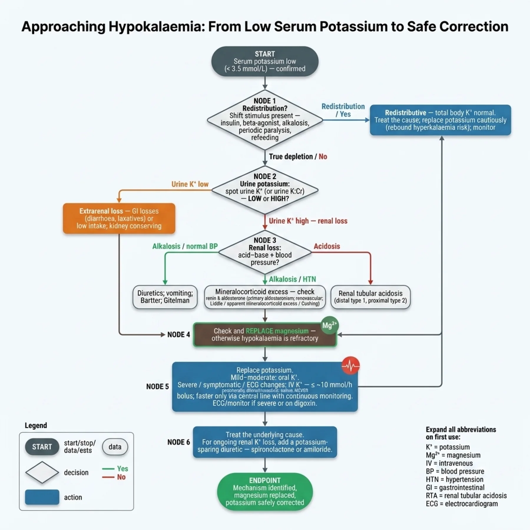
Replacement is governed by safety as much as by the deficit. Oral potassium (potassium chloride) is preferred for mild to moderate depletion because it is safer and the gut moderates absorption; intravenous potassium is reserved for severe hypokalaemia, symptomatic or ECG-positive cases, and patients who cannot take it orally. The intravenous route carries real hazard and strict rules: a controlled infusion rate (broadly up to about 10 mmol/hour peripherally, higher only with central access and cardiac monitoring), dilution in saline rather than dextrose (dextrose triggers insulin, which shifts potassium into cells and worsens the hypokalaemia), continuous cardiac monitoring at higher rates, and never a bolus, which can be fatal. The deficit is hard to estimate precisely — a rough guide is that each 0.3 mmol/L fall represents roughly 100 mmol of total deficit — so replacement is titrated against repeated measurement. Magnesium is replaced if low. The cause is treated — stopping or changing a diuretic, addressing mineralocorticoid excess with spironolactone or amiloride — and in redistributive hypokalaemia the cause is treated with cautious replacement to avoid the rebound. Throughout, the contrast with hyperkalaemia's emergency is instructive: hypokalaemia is usually corrected steadily and safely, with the intravenous route the main source of iatrogenic danger.
— Where the mechanisms meet the bedside
The unifying thread is that the plasma potassium is a small, shiftable window onto a large store governed by two balances and one indispensable cofactor. Diagnosis asks, in order: is this a shift or a true deficit; if a deficit, is the loss renal or extrarenal; if renal, what does the acid-base and blood-pressure pattern say; and is the magnesium low? Treatment then replaces what is truly missing, by the safest adequate route, replaces magnesium, treats the cause, and resists over-replacing a shift. Two errors recur and are worth pre-empting: over-replacing redistributive hypokalaemia into rebound hyperkalaemia, and chasing a hypokalaemia that will not correct without the magnesium it depends on. Get the mechanism right first, and the treatment becomes both effective and safe.
| 04 | PHASE A · LEVEL 4 · ORIENTATION & KNOWLEDGE Reference Tables |
Table 7.1 — Mechanisms of hypokalaemia
| Mechanism | Detail |
| Redistribution (shift in) | Insulin, beta-agonists, alkalosis, periodic paralysis, refeeding — total body normal |
| True depletion — renal | Diuretics, mineralocorticoid excess, Bartter/Gitelman, RTA, hypomagnesaemia |
| True depletion — extrarenal | Gastrointestinal (diarrhoea); low intake |
| Key first question | Shift or true deficit? — they are managed differently |
Table 7.2 — Potassium balance
| System | Detail |
| Internal balance | Na-K-ATPase shifts K between cells and plasma (insulin, beta, pH) |
| External balance | Total-body K — renal excretion (main) and GI |
| Renal secretion | Distal nephron: aldosterone, distal Na delivery/flow, lumen-negative voltage, ROMK |
| Plasma fraction | ~2% of total body K — small shifts move it a lot |
Table 7.3 — Localising depletion: urine potassium
| Urine potassium | Interpretation |
| Low (< ~15–20 mmol/day; low K/Cr) | Extrarenal loss (GI) or low intake — kidney conserving |
| High (inappropriate) | Renal loss — classify further by acid-base and BP |
| Confirm | Pseudohypokalaemia (high WBC in vitro) is rare |
Table 7.4 — Classifying renal loss
| Pattern | Causes |
| Alkalosis, normal/low BP | Diuretics, vomiting (2° hyperaldosteronism), Bartter, Gitelman |
| Alkalosis, hypertension | Mineralocorticoid excess: Conn's, Cushing's, Liddle, renovascular |
| Metabolic acidosis | Renal tubular acidosis (type 1/2), treated DKA, ureteral diversion |
| Always check | Magnesium — hypomagnesaemia causes renal K wasting |
Table 7.5 — ECG and arrhythmia
| Feature | Detail |
| ECG changes | T-wave flattening/inversion, ST depression, U waves, QT/QU prolongation |
| Arrhythmia | Ventricular arrhythmias in severe hypokalaemia |
| Digoxin | Hypokalaemia potentiates digoxin toxicity — dangerous combination |
| Urgency | ECG changes/arrhythmia/digoxin → prompt monitored correction |
Table 7.6 — Treatment
| Element | Detail |
| Oral KCl | Preferred for mild–moderate (safer) |
| Intravenous KCl | Severe/symptomatic/non-oral — ≤ ~10 mmol/h peripherally; in saline, NOT dextrose; never bolus |
| Magnesium | Replace if low — otherwise hypokalaemia is refractory |
| Redistribution | Treat the cause; replace cautiously (rebound hyperkalaemia risk) |
| Cause / ongoing loss | Treat; spironolactone/amiloride for renal loss or mineralocorticoid excess |
| 05 | PHASE B · LEVEL 5 · VISUALISE & MAP Imaging & Flowchart Specifications |
| 06 | PHASE B · LEVEL 6 · VISUALISE & MAP Concept Maps |
Each chain runs from physiology to a named bedside action; read the arrows as “leads to.”
The two balances. Potassium ~98% intracellular → internal balance (Na-K-ATPase shifts) buffers plasma; external balance (renal/GI) sets total body → ACTION: ask whether a low potassium is a shift or a true deficit before treating.
Renal secretion. Distal nephron secretes K driven by aldosterone, distal Na delivery/flow, and lumen-negative voltage via ROMK → disorders of these waste K → ACTION: classify renal loss by acid-base and BP (aldosterone-driven states).
Magnesium–potassium. Magnesium inhibits ROMK; hypomagnesaemia lifts the brake → unchecked K secretion → refractory hypokalaemia → ACTION: check and replace magnesium in any persistent hypokalaemia.
Redistribution vs depletion. Shift into cells (total body normal) vs true loss → shift reverses when the stimulus resolves → ACTION: treat redistribution's cause and replace cautiously to avoid rebound hyperkalaemia.
ECG / arrhythmia. Low potassium → altered membrane excitability → U waves, QT/QU prolongation, ventricular arrhythmia (worse with digoxin) → ACTION: let ECG changes, arrhythmia, and digoxin set the urgency of correction.
| 07 | PHASE B · LEVEL 7 · VISUALISE & MAP Decision Pathways |
| R1 | IF a patient is hypokalaemic, THEN first decide whether it is redistribution (a shift) or true depletion — they are managed differently. |
| R2 | IF the hypokalaemia is redistributive, THEN treat the cause and replace cautiously — over-replacement causes rebound hyperkalaemia when the shift reverses. |
| R3 | IF the hypokalaemia is depletional, THEN use the urine potassium — low means extrarenal/low intake, high means renal loss. |
| R4 | IF the loss is renal, THEN classify by acid-base and blood pressure (alkalosis ± hypertension, or acidosis) and investigate accordingly. |
| R5 | IF hypokalaemia is refractory, THEN check and replace magnesium — hypomagnesaemia causes renal potassium wasting. |
| R6 | IF there are ECG changes, arrhythmia, or digoxin therapy, THEN correct promptly with monitoring — the arrhythmia risk is high. |
| R7 | IF replacing intravenously, THEN limit the rate (~10 mmol/h peripherally), use saline not dextrose, monitor cardiac rhythm at higher rates, and never bolus. |
| R8 | IF there is ongoing renal loss or mineralocorticoid excess, THEN treat the cause and consider a potassium-sparing agent (spironolactone or amiloride). |
| 08 | PHASE C · LEVEL 8 · CLINICAL REASONING Clinical Cases |
| CASE 1 | IT WON'T COME UP The forgotten magnesium Refractory hypokalaemia |
Presentation
A patient on long-term diuretics has a persistent potassium of 2.9 despite repeated potassium replacement over two days. The team keeps giving more potassium with little effect.
❖ Pause and reflect Why won't the potassium correct despite replacement? |
Analysis
The likely culprit is hypomagnesaemia. Diuretics waste both potassium and magnesium, and a low magnesium lifts the inhibition of the ROMK channel so the kidney secretes potassium unchecked — it excretes the potassium being given. Until the magnesium is replaced, the hypokalaemia is refractory, however much potassium is administered. This is the commonest reason hypokalaemia 'refuses' to respond, and checking the magnesium should have been an early step.
Plan
Check and replace the magnesium, then continue potassium replacement — the potassium will now correct. Review the diuretic as the shared cause of both deficits.
Teaching point
Refractory hypokalaemia is hypomagnesaemia until proven otherwise — replace magnesium or the potassium will not correct.
Cross-reference
Exercises rule R5; the magnesium-potassium concept map; Figure 7.3; Tables 7.4 and 7.6.
| CASE 2 | POTASSIUM IN HIDING Don't over-replace a shift Redistributive hypokalaemia |
Presentation
A young patient presents with acute flaccid weakness and a potassium of 2.2 during an episode of hypokalaemic periodic paralysis. The team plans aggressive intravenous potassium to normalise the level quickly.
❖ Pause and reflect Is aggressive replacement the right approach to this hypokalaemia? |
Analysis
This is redistributive hypokalaemia — the potassium has shifted into cells, not left the body, so total body potassium is normal. Aggressive replacement risks rebound hyperkalaemia when the shift reverses and the potassium floods back into the plasma, which can be dangerous. The correct approach is cautious replacement to relieve the acute weakness and arrhythmia risk, with the expectation that the potassium will return on its own as the episode resolves — and close monitoring to catch the rebound.
Plan
Replace potassium cautiously to address the acute danger, monitor closely for rebound hyperkalaemia as the shift reverses, and treat the underlying paralysis. Do not aggressively replace as if this were true depletion.
Teaching point
Redistributive hypokalaemia is potassium in hiding, not lost — replace cautiously to avoid rebound hyperkalaemia.
Cross-reference
Exercises rules R1 and R2; the redistribution concept map; Tables 7.1 and 7.6.
| CASE 3 | HYPERTENSION AND LOW POTASSIUM Acid-base and blood pressure Mineralocorticoid excess |
Presentation
A hypertensive patient has persistent hypokalaemia with a metabolic alkalosis, a high urine potassium, and no diuretic use. The team is unsure where the renal potassium loss is coming from.
❖ Pause and reflect What does the combination of hypertension, hypokalaemia, and alkalosis point to? |
Analysis
This is the classic triad of mineralocorticoid excess. The high urine potassium confirms renal wasting; the metabolic alkalosis and — critically — the hypertension point to an excess of mineralocorticoid activity driving distal sodium reabsorption and potassium secretion. The differential is primary hyperaldosteronism (Conn's syndrome), Cushing's syndrome, Liddle syndrome, and renovascular disease, distinguished by the renin and aldosterone. The two-axis classification — alkalosis with hypertension — has narrowed a long list to this group.
Plan
Measure renin and aldosterone to characterise the mineralocorticoid excess, investigate for the specific cause, replace potassium and magnesium, and use a potassium-sparing agent (spironolactone or amiloride) for the ongoing renal loss while the cause is addressed.
Teaching point
Hypertension with hypokalaemia and alkalosis and renal potassium loss points to mineralocorticoid excess — check renin and aldosterone.
Cross-reference
Exercises rules R4 and R8; the renal-secretion concept map; Figure 7.2; Table 7.4.
| CASE 4 | GETTING THE REPLACEMENT RIGHT Route, rate, and the saline detail Safe potassium repletion |
Presentation
A patient with severe symptomatic hypokalaemia (potassium 2.3, U waves on the ECG) needs intravenous potassium. A trainee prepares to give it rapidly in a dextrose solution through a peripheral line.
❖ Pause and reflect What is wrong with this replacement plan? |
Analysis
Two errors. First, the rate: peripheral intravenous potassium should run at no more than about 10 mmol/hour, with higher rates only via central access and cardiac monitoring, and never as a bolus, which can be fatal. Second, the diluent: potassium should be given in saline, not dextrose, because dextrose triggers insulin secretion, which shifts potassium into cells and transiently worsens the hypokalaemia. With ECG changes present, continuous cardiac monitoring is also needed. The severity here justifies the intravenous route, but it must be given safely.
Plan
Give intravenous potassium in saline at a controlled rate (~10 mmol/hour peripherally, higher only centrally with monitoring), never as a bolus, with continuous ECG monitoring, and check and replace magnesium. Titrate against repeated potassium measurement.
Teaching point
Intravenous potassium: controlled rate, in saline not dextrose, never a bolus, with monitoring when severe — the route's danger is iatrogenic.
Cross-reference
Exercises rules R6 and R7; the ECG concept map; Tables 7.5 and 7.6.
| 09 | PHASE C · LEVEL 9 · CLINICAL REASONING Clinical Implications |
One triad per mechanism the narrative exposed: the physiology, why it matters, and the bedside move.
MECHANISM Potassium is overwhelmingly intracellular, so plasma reflects only a small, shiftable fraction. |
WHY IT MATTERS A low potassium can be a shift into cells or a true total-body deficit. |
ACTION Decide shift versus deficit before treating. |
MECHANISM The distal nephron secretes potassium under aldosterone, flow, and the lumen-negative voltage. |
WHY IT MATTERS Disorders of these mechanisms waste potassium renally. |
ACTION Classify renal loss by acid-base status and blood pressure. |
MECHANISM Magnesium inhibits the ROMK channel, and hypomagnesaemia lifts that inhibition. |
WHY IT MATTERS The kidney then wastes potassium and the hypokalaemia is refractory to potassium alone. |
ACTION Check and replace magnesium in any persistent hypokalaemia. |
MECHANISM In redistribution the potassium has shifted into cells, not left the body. |
WHY IT MATTERS It returns to the plasma when the stimulus resolves, so over-replacement causes rebound. |
ACTION Treat the cause and replace cautiously. |
MECHANISM Hypokalaemia alters cardiac membrane excitability and potentiates digoxin. |
WHY IT MATTERS It predisposes to ventricular arrhythmia, especially with digoxin or cardiac disease. |
ACTION Let ECG changes, arrhythmia, and digoxin set the urgency of correction. |
| 10 | PHASE C · LEVEL 10 · CLINICAL REASONING Clinical Pearls |
| Potassium is ~98% intracellular — plasma is a thin, shiftable window. | Internal balance: Na-K-ATPase shifts (insulin, beta, alkalosis → in). |
| External balance: renal (main) and GI loss set total body potassium. | First question: redistribution (shift) or true depletion? |
| Redistribution reverses — over-replacement → rebound hyperkalaemia. | Depletion: urine K low = extrarenal/low intake; high = renal. |
| Renal loss + alkalosis + normal BP: diuretics, vomiting, Bartter, Gitelman. | Renal loss + alkalosis + HTN: mineralocorticoid excess (check renin/aldosterone). |
| Renal loss + acidosis: renal tubular acidosis, treated DKA. | Hypomagnesaemia → renal K wasting → refractory hypokalaemia. |
| ALWAYS replace magnesium in refractory hypokalaemia. | ECG: U waves, T flattening, ST depression, QT/QU prolongation. |
| Hypokalaemia potentiates digoxin toxicity — dangerous combination. | Oral KCl preferred (mild–moderate); IV for severe/symptomatic/non-oral. |
| IV K: ≤ ~10 mmol/h peripheral; in saline NOT dextrose; never bolus; monitor. | Treat the cause; spironolactone/amiloride for ongoing renal loss. |
| 11 | PHASE D · LEVEL 11 · SAFETY & EVIDENCE Red Flags & Never-Do |
Panel A — Red flags
| ▲ | Hypokalaemia that won't correct despite replacement — check and replace magnesium. |
| ▲ | Severe hypokalaemia in a patient on digoxin — high arrhythmia risk; correct promptly with monitoring. |
| ▲ | Aggressive replacement of redistributive hypokalaemia (periodic paralysis) — rebound hyperkalaemia risk; replace cautiously. |
| ▲ | Hypertension with hypokalaemia and alkalosis — mineralocorticoid excess; check renin and aldosterone. |
| ▲ | ECG U waves / QT-QU prolongation with severe hypokalaemia — arrhythmia risk; urgent monitored correction. |
Panel B — Never do
| ✖ NEVER — chase a refractory hypokalaemia without checking the magnesium. |
| ✖ NEVER — aggressively replace a clearly redistributive hypokalaemia. |
| ✖ NEVER — give intravenous potassium as a bolus, or in dextrose, or uncontrolled. |
| ✖ NEVER — ignore digoxin therapy when correcting hypokalaemia. |
| 12 | PHASE D · LEVEL 12 · SAFETY & EVIDENCE Common Pitfalls |
Pitfall 1 — Forgetting magnesium
| ✖ | WRONG Giving ever-more potassium to a refractory hypokalaemia. |
| ✓ | RIGHT Checking and replacing the magnesium. |
| ✉ | WHY Hypomagnesaemia causes renal potassium wasting that defeats replacement. |
Pitfall 2 — Over-replacing a shift
| ✖ | WRONG Aggressively replacing redistributive hypokalaemia (e.g. periodic paralysis). |
| ✓ | RIGHT Replacing cautiously and watching for rebound. |
| ✉ | WHY The potassium returns to plasma when the shift reverses, causing hyperkalaemia. |
Pitfall 3 — Skipping the classification
| ✖ | WRONG Replacing potassium without finding the source of renal loss. |
| ✓ | RIGHT Using urine potassium, acid-base status, and blood pressure to classify. |
| ✉ | WHY The cause (e.g. mineralocorticoid excess) needs specific treatment. |
Pitfall 4 — Unsafe IV potassium
| ✖ | WRONG Giving rapid intravenous potassium in dextrose. |
| ✓ | RIGHT Controlled rate in saline, never a bolus, with monitoring. |
| ✉ | WHY Boluses can be fatal and dextrose worsens the hypokalaemia via insulin. |
Pitfall 5 — Ignoring digoxin
| ✖ | WRONG Treating hypokalaemia leisurely in a digoxin-treated patient. |
| ✓ | RIGHT Correcting promptly with monitoring. |
| ✉ | WHY Hypokalaemia potentiates digoxin toxicity and arrhythmia. |
| 13 | PHASE D · LEVEL 13 · SAFETY & EVIDENCE Evidence Grading |
GRADE A HIGH CONFIDENCE The effect is real and the estimate is stable. RCTs at low risk of bias; multiple concordant prospective cohorts; meta-analyses. |
GRADE B MODERATE CONFIDENCE The effect is likely real but may shift with new data. Observational studies, registries, mechanistic human studies. |
GRADE C LOW CONFIDENCE Rests on physiology, reasoning, or consensus rather than outcomes. Pathophysiological reasoning; extrapolation; consensus without outcomes. |
Graded statements (by evidence type)
| Statement | Grade | Basis (evidence type) |
| Plasma potassium reflects a small fraction of total body potassium. | A | Established physiology |
| Insulin, beta-agonists, and alkalosis shift potassium into cells. | A | Established physiology |
| Hypomagnesaemia causes renal potassium wasting and refractory hypokalaemia. | A | Established physiology and clinical data |
| The urine potassium localises depletion as renal or extrarenal. | A | Established physiology |
| Hypokalaemia potentiates digoxin toxicity. | A | Established pharmacology |
| Over-replacement of redistributive hypokalaemia causes rebound hyperkalaemia. | B | Clinical observation |
| Intravenous potassium boluses are dangerous and contraindicated. | A | Pharmacology and safety data |
| 14 | PHASE E · LEVEL 14 · PATIENT DECISIONS Absolute Risk in Natural Frequency |
Natural-frequency estimates for orientation, from hypokalaemia management; they vary with severity and comorbidity. They convey the size of the decisions, expressed per 100 comparable patients.
| Per 100 patients… | Outcome | Roughly how many | See |
| Refractory hypokalaemia given magnesium | Finally correct the potassium | Most — if magnesium was the missing factor | L13 row 3 |
| Redistributive hypokalaemia over-replaced | Develop rebound hyperkalaemia | More than the cautiously replaced | L13 row 6 |
| Severe hypokalaemia on digoxin | Suffer an arrhythmia | More than those without digoxin | L13 row 5 |
| Given an intravenous potassium bolus | Suffer a fatal arrhythmia | An unacceptable number — hence never bolus | L13 row 7 |
★ How to read these Read these as orientation, not promises; outcomes vary with severity and comorbidity. The stable signals: magnesium rescues refractory hypokalaemia, over-replacing a shift rebounds, digoxin amplifies the danger, and bolus potassium kills. Communicate them as people out of 100, not as a hazard ratio. |
| 17 | PHASE F · LEVEL 17 · APPLY & TEST Documentation Templates |
Paste-ready notes. Tick the boxes that apply and delete the rest; make the mechanism, the magnesium, and the safe route explicit.
Template 1 — Hypokalaemia assessment
Template 2 — Repletion plan
| 18 | PHASE F · LEVEL 18 · APPLY & TEST Cheat Sheet |
| K ~98% intracellular — plasma is a thin, shiftable window. | Internal: Na-K-ATPase (insulin, beta, alkalosis → in). |
| External: renal (main) + GI. | First Q: shift (redistribution) or true deficit? |
| Redistribution reverses → over-replacement = rebound hyperkalaemia. | Depletion: urine K low = extrarenal/low intake; high = renal. |
| Renal + alkalosis + normal BP: diuretics, vomiting, Bartter, Gitelman. | Renal + alkalosis + HTN: mineralocorticoid excess (renin/aldosterone). |
| Renal + acidosis: RTA, treated DKA. | Low magnesium → renal K wasting → refractory hypokalaemia. |
| ALWAYS replace magnesium if refractory. | ECG: U waves, T flattening, ST depression, QT/QU prolongation. |
| Hypokalaemia + digoxin = dangerous. | Oral KCl preferred; IV for severe/symptomatic/non-oral. |
| IV K: ≤ ~10 mmol/h, in saline NOT dextrose, never bolus, monitor. | Treat cause; spironolactone/amiloride for ongoing renal loss. |
| 19 | PHASE F · LEVEL 19 · APPLY & TEST Flashcards |
| CARD 1 | Q. Why does the plasma potassium misrepresent total body potassium? A. Because about 98% of potassium is intracellular, the plasma holds only a small fraction, so a shift across the membrane or a modest total deficit moves the plasma level a great deal. DETAILED. Internal balance buffers plasma without changing total body potassium. CLINICAL. Decide shift versus deficit before treating. |
| CARD 2 | Q. What drives potassium into cells? A. Activation of the sodium-potassium pump by insulin and beta-adrenergic stimulation, and alkalosis (potassium taken up in exchange for hydrogen ions). DETAILED. These cause redistributive hypokalaemia without true loss. CLINICAL. Treat the cause and replace cautiously to avoid rebound. |
| CARD 3 | Q. How does the urine potassium localise depletion? A. A low urine potassium (under ~15–20 mmol/day) indicates the kidney is conserving — extrarenal loss or low intake; a high urine potassium indicates inappropriate renal wasting. DETAILED. It splits depletion into extrarenal and renal groups. CLINICAL. Use it as the second diagnostic step. |
| CARD 4 | Q. How is renal potassium loss classified? A. By acid-base status and blood pressure: alkalosis with normal/low BP (diuretics, vomiting, Bartter, Gitelman), alkalosis with hypertension (mineralocorticoid excess), and acidosis (renal tubular acidosis). DETAILED. It turns a long list into a short scheme. CLINICAL. Use acid-base and blood pressure to narrow the cause. |
| CARD 5 | Q. Why does hypomagnesaemia cause refractory hypokalaemia? A. Magnesium normally inhibits the ROMK channel; when magnesium is low, the inhibition is lifted and the channel secretes potassium unchecked, so the kidney wastes the potassium being replaced. DETAILED. The hypokalaemia will not correct until magnesium is replaced. CLINICAL. Check and replace magnesium in persistent hypokalaemia. |
| CARD 6 | Q. What are the ECG features and the key drug interaction? A. T-wave flattening and inversion, ST depression, U waves, and QT-QU prolongation, with a risk of ventricular arrhythmia — sharply amplified by digoxin, which hypokalaemia potentiates. DETAILED. ECG changes and digoxin raise the urgency. CLINICAL. Correct promptly with monitoring when these are present. |
| CARD 7 | Q. How is potassium replaced safely? A. Oral potassium for mild to moderate depletion; intravenous for severe, symptomatic, or non-oral cases at a controlled rate (~10 mmol/hour peripherally), diluted in saline not dextrose, never as a bolus, with monitoring. DETAILED. Dextrose triggers insulin and worsens the hypokalaemia. CLINICAL. Use the safest adequate route and replace magnesium too. |
| CARD 8 | Q. Why must redistributive hypokalaemia be replaced cautiously? A. Because the potassium has shifted into cells, not left the body; when the stimulus resolves it returns to the plasma, so aggressive replacement causes rebound hyperkalaemia. DETAILED. Total body potassium is normal in a pure shift. CLINICAL. Treat the cause and replace cautiously, watching for rebound. |
| 20 | PHASE F · LEVEL 20 · APPLY & TEST One-Minute Preceptor |
| SCENE 1 | The intern chasing the potassium |
GET A COMMITMENT. “You've given a lot of potassium and it's still 2.9 — what's going on?”
PROBE FOR EVIDENCE. “It just won't come up” — ask: “Have you checked the magnesium, and what does low magnesium do to renal potassium handling?”
TEACH A GENERAL RULE. Hypomagnesaemia lifts the brake on the ROMK channel, so the kidney wastes the potassium you give — refractory hypokalaemia is hypomagnesaemia until proven otherwise.
REINFORCE WHAT WAS RIGHT. Recognising the inadequate response was correct.
CORRECT A MISTAKE. Check and replace the magnesium, then continue potassium.
| SCENE 2 | The resident over-replacing a shift |
GET A COMMITMENT. “You're giving aggressive potassium for this periodic paralysis — why?”
PROBE FOR EVIDENCE. “The potassium is 2.2” — ask: “Is the potassium lost from the body or shifted into cells, and what happens when the episode resolves?”
TEACH A GENERAL RULE. In redistribution the potassium is hiding inside cells, not lost; it returns when the shift reverses, so aggressive replacement causes rebound hyperkalaemia.
REINFORCE WHAT WAS RIGHT. Addressing the acute danger was reasonable.
CORRECT A MISTAKE. Replace cautiously and watch for rebound.
| 22 | PHASE F · LEVEL 22 · APPLY & TEST Board-Style Questions |
| Q 01 | Why does a small total-body potassium change move the plasma level so much? |
| A | Potassium is mostly extracellular |
| B | Potassium is ~98% intracellular, so plasma reflects a tiny, shiftable fraction |
| C | The kidney cannot excrete potassium |
| D | Plasma potassium is fixed |
Rationale With most potassium intracellular, the plasma is a thin window, so shifts and modest deficits move it markedly (Figure 7.1, rule R1). A, C, and D are incorrect. |
| Q 02 | The first step in evaluating hypokalaemia is to determine whether it is: |
| A | Renal or extrarenal |
| B | Redistribution (a shift) or true depletion |
| C | Acute or chronic |
| D | Symptomatic or not |
Rationale Shift versus deficit is the first and most consequential question because they are managed differently (rule R1, Table 7.1). A is the next step in depletion; C and D inform urgency, not mechanism. |
| Q 03 | Why won't a hypokalaemia correct despite repeated potassium replacement? |
| A | Too little potassium given |
| B | Coexisting hypomagnesaemia causing renal potassium wasting |
| C | The potassium is being measured wrong |
| D | It is redistributive |
Rationale Low magnesium lifts ROMK inhibition so the kidney wastes the replaced potassium — the classic refractory hypokalaemia (case 1, Figure 7.3, rule R5). A is unlikely with repeated dosing; C and D don't fit refractoriness to replacement. |
| Q 04 | A hypertensive patient has hypokalaemia, metabolic alkalosis, and a high urine potassium. This suggests: |
| A | Gastrointestinal loss |
| B | Mineralocorticoid excess — check renin and aldosterone |
| C | Redistribution |
| D | Renal tubular acidosis |
Rationale Renal wasting with alkalosis and hypertension is the triad of mineralocorticoid excess (case 3, Table 7.4, rule R4). A gives low urine K; C is a shift; D causes acidosis. |
| Q 05 | How should redistributive hypokalaemia (e.g. periodic paralysis) be treated? |
| A | Aggressive potassium replacement |
| B | Cautious replacement and treating the cause, watching for rebound |
| C | No treatment ever |
| D | Intravenous bolus potassium |
Rationale The potassium is shifted, not lost, and returns when the shift reverses, so over-replacement rebounds (case 2, rule R2). A and D cause rebound/harm; C ignores acute danger. |
| Q 06 | Which is correct for intravenous potassium replacement? |
| A | Bolus is acceptable if severe |
| B | Controlled rate (~10 mmol/h peripheral), in saline not dextrose, never a bolus |
| C | Always use dextrose as the diluent |
| D | No monitoring is needed |
Rationale Safe IV potassium is rate-limited, in saline (dextrose triggers insulin and worsens it), never bolused, and monitored when severe (case 4, rule R7). A, C, and D are dangerous. |
| Q 07 | Why is hypokalaemia especially dangerous in a patient on digoxin? |
| A | It has no interaction |
| B | Hypokalaemia potentiates digoxin toxicity and arrhythmia |
| C | It lowers digoxin levels |
| D | Digoxin raises potassium |
Rationale Hypokalaemia potentiates digoxin toxicity, raising arrhythmia risk and the urgency of correction (Table 7.5, rule R6). A, C, and D are incorrect. |
| Q 08 | A low urine potassium in a hypokalaemic patient indicates: |
| A | Renal wasting |
| B | Extrarenal loss or low intake — the kidney is conserving |
| C | Mineralocorticoid excess |
| D | Diuretic use |
Rationale A low urine potassium means appropriate renal conservation, pointing to gastrointestinal loss or low intake (Table 7.3, rule R3). A, C, and D cause renal wasting (high urine K). |
| Q 09 | Across 100 patients with refractory hypokalaemia in whom magnesium was the missing factor, replacing magnesium: |
| A | Helps few |
| B | Allows the potassium to correct in most |
| C | Has no effect |
| D | Worsens the potassium |
Rationale When hypomagnesaemia is the cause of refractoriness, replacing it allows the potassium to correct (L14, L13 row 3). A, C, and D contradict the mechanism. |