Chapter Preamble
Signals declared
Sig-D — Diagnostic (primary). Assess extracellular volume and plasma tonicity as two separate axes, integrating clinical signs and adjuncts.
Sig-M — Mechanistic (strong). The fluid compartments, osmosis and tonicity, and the foundational distinction between sodium content (volume) and sodium concentration (water) that organises the whole volume.
Levels populated and omitted
Populated (17): L1–L13, L18–L20, L22. The mechanistic signal fires the concept maps (L6) and the mechanism-to-action triads (L9); the diagnostic signal drives the tables, rules, cases, pitfalls, and board items.
L14 absolute-risk — omitted. No Sig-T/E/V; this foundational chapter explains physiology and assessment rather than quantifying treatment outcomes.
L15 / L16 preference-sensitive map and SDM scripts — omitted. No Sig-E; understanding compartments and assessing volume is foundational knowledge — and electrolyte care, throughout this volume, is effective care rather than a values-driven choice.
L17 documentation templates — omitted. No Sig-P/T; the therapies that act on these compartments are built in the chapters that follow.
| 01 | PHASE A · LEVEL 1 · ORIENTATION & KNOWLEDGE Learning Objectives |
By the end of this chapter you should be able to:
Describe the body fluid compartments and their characteristic solutes.
Distinguish osmolality from tonicity and explain why effective and ineffective osmoles differ.
Explain why sodium content determines extracellular volume while water balance determines sodium concentration.
Separate the assessment of extracellular volume from the assessment of plasma tonicity.
Assess extracellular volume status by integrating clinical signs rather than relying on any single one.
Recognise discordance between intravascular and interstitial volume.
Use adjuncts — urine sodium, laboratory clues, and point-of-care ultrasound — to refine volume assessment.
Distinguish volume status from volume responsiveness.
| 02 | PHASE A · LEVEL 2 · ORIENTATION & KNOWLEDGE Executive Summary |
Total body water is about 60% of body weight in men and 50% in women, lower in the elderly and the obese.
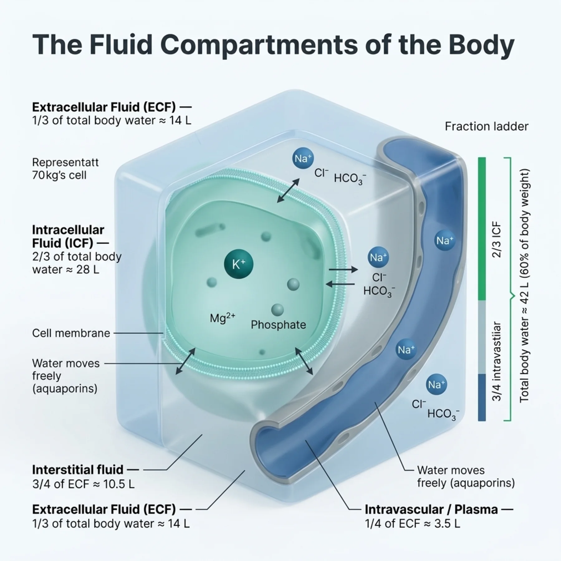
Two-thirds of body water is intracellular and one-third extracellular; the extracellular fluid is three-quarters interstitial and one-quarter intravascular.
Potassium, magnesium, and phosphate dominate the intracellular fluid, while sodium, chloride, and bicarbonate dominate the extracellular fluid.
Plasma osmolality is the total solute concentration; tonicity is the effective osmolality — only the osmoles that do not cross membranes freely.
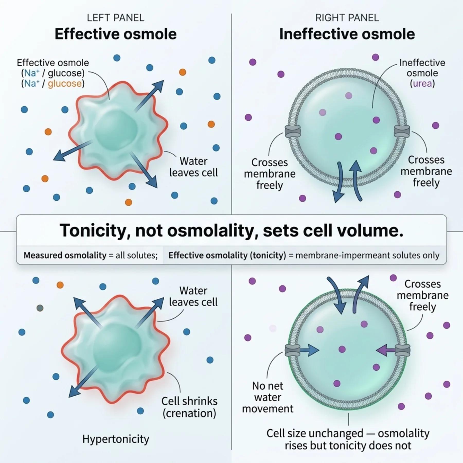
Effective osmoles such as sodium and glucose draw water across membranes; ineffective osmoles such as urea and ethanol cross freely and cause no sustained shift.
Tonicity, not osmolality, determines cell volume, so it is tonicity that the dysnatraemias threaten.
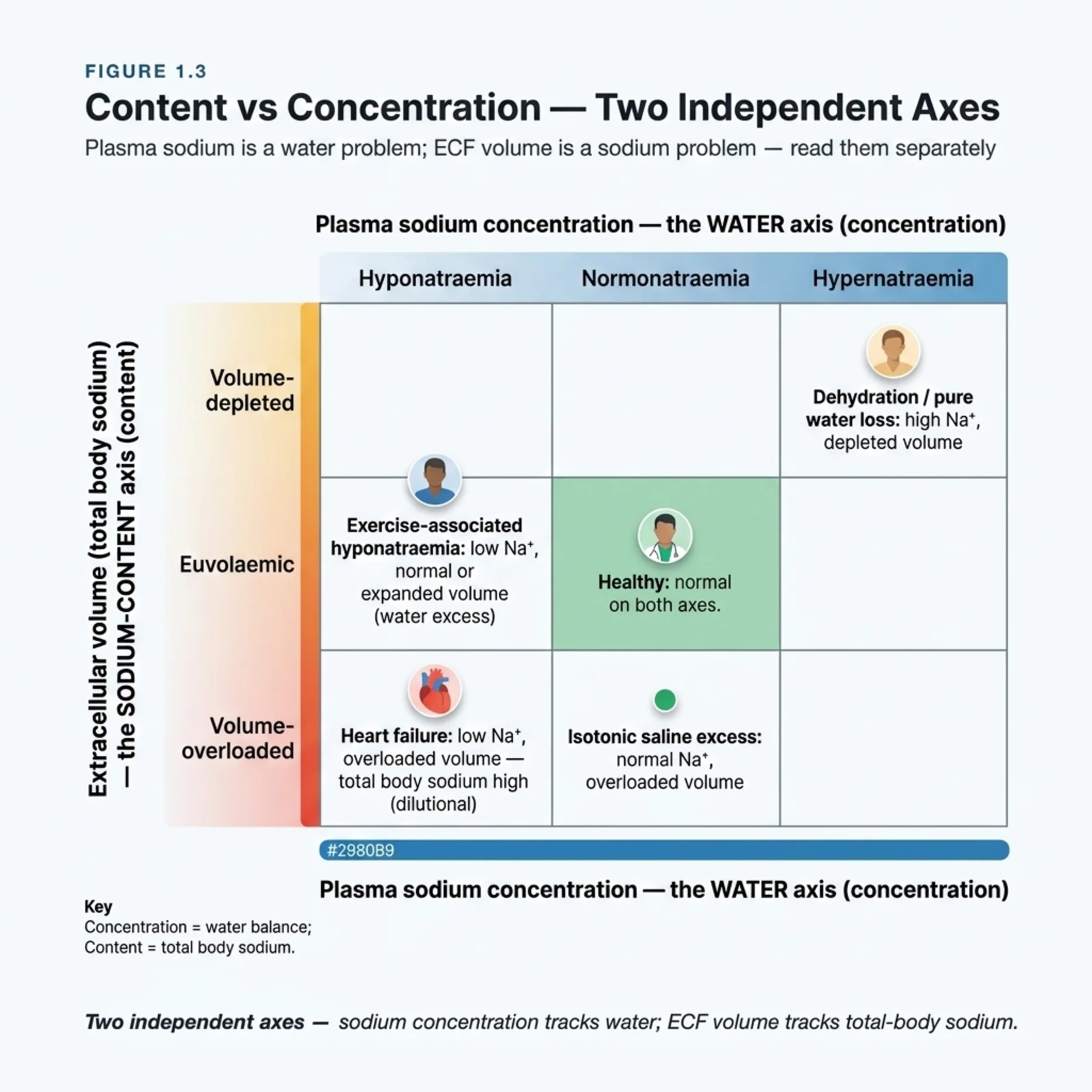
The foundational distinction of the whole volume: sodium content determines extracellular volume, while water balance determines sodium concentration.
Disorders of extracellular volume — depletion and overload — are sodium problems; the dysnatraemias are water problems.
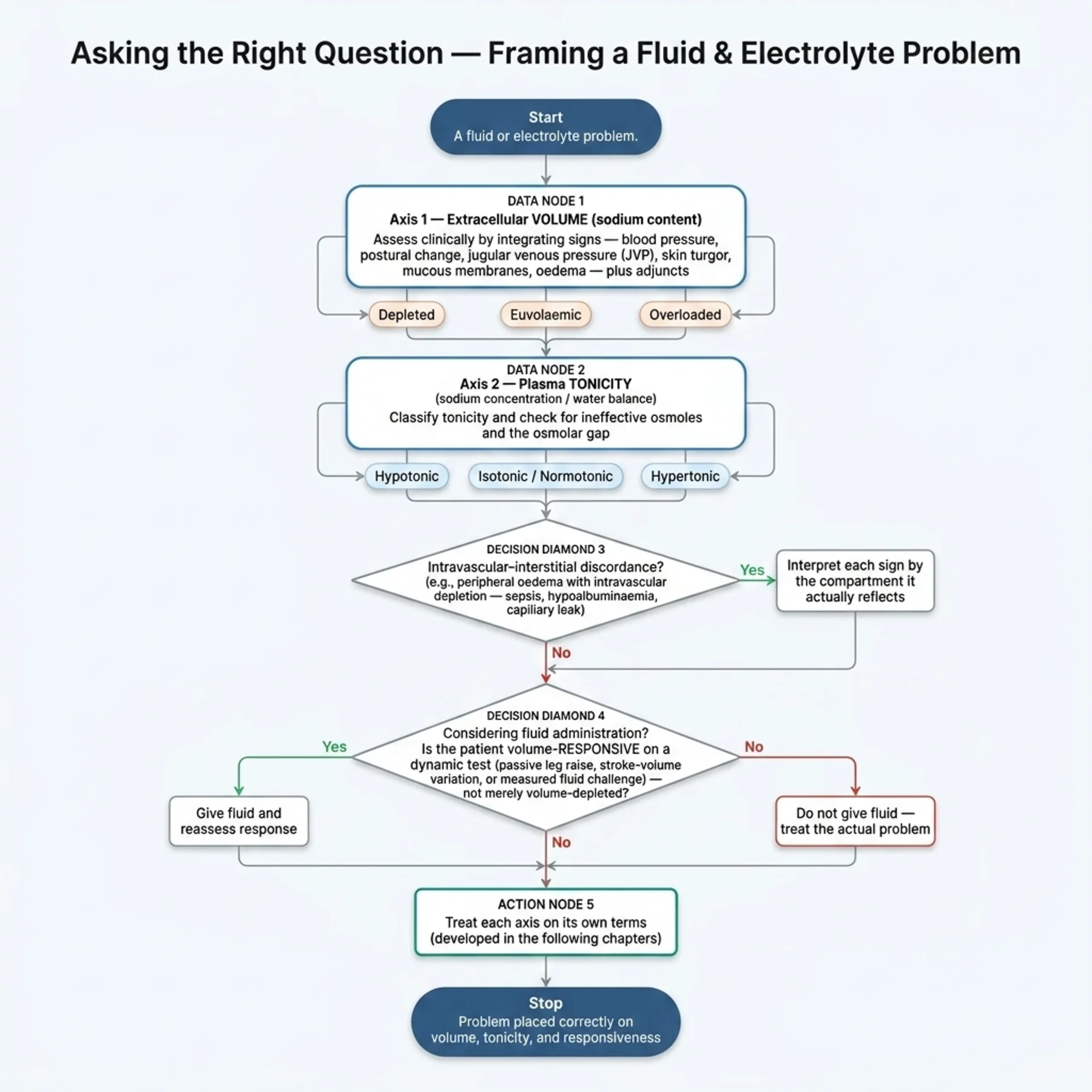
Volume status and plasma tonicity are therefore two separate axes, assessed separately.
Clinical signs of extracellular volume — turgor, mucous membranes, jugular venous pressure, oedema, postural change, urine output, weight — are individually insensitive and must be integrated.
Intravascular and interstitial volume can diverge, as in hypoalbuminaemia or capillary leak with oedema yet intravascular depletion.
Adjuncts — urine sodium, the urea-to-creatinine ratio, haematocrit, and point-of-care ultrasound — refine the clinical picture.
Volume status is not the same as volume responsiveness: whether fluid will help is a dynamic question, not a static one.
Getting these distinctions right at the outset is what makes the rest of the volume coherent.
| 03 | PHASE A · LEVEL 3 · ORIENTATION & KNOWLEDGE Main Narrative |
Almost every error in fluid and electrolyte medicine traces back to a confusion of two ideas: how much salt water the body holds, and how concentrated that water is. The first is extracellular volume, governed by sodium content; the second is tonicity, governed by water balance and reflected in the sodium concentration. They are different axes, assessed differently and corrected differently, and the patient can be abnormal on either, both, or neither. This opening chapter builds the compartments, the physics of water movement, and that central distinction, because everything that follows depends on it.
— The compartments
Body water is distributed in nested compartments. Total body water is roughly 60% of body weight in men and 50% in women — lower in the elderly and the obese, because fat holds little water — and it divides into two-thirds intracellular and one-third extracellular. The extracellular fluid divides again into the interstitial space, about three-quarters, and the intravascular plasma, about one-quarter. Each compartment has a characteristic ionic composition: the intracellular fluid is rich in potassium, magnesium, and phosphate, held there by cellular pumps and proteins, while the extracellular fluid is dominated by sodium, chloride, and bicarbonate. Sodium is the principal extracellular cation, and because of that it is sodium that determines how much water sits in the extracellular space — a point that becomes the organising idea of the chapter.
— Osmolality, tonicity, and why they differ
Water moves across cell membranes by osmosis, from lower to higher solute concentration, until concentrations equalise. But not all solutes pull water the same way, and this is where osmolality and tonicity part company. Osmolality is the total concentration of all solutes, measured or calculated as roughly twice the sodium plus glucose plus urea, and normally 275 to 295. Tonicity — the effective osmolality — counts only the solutes that cannot cross the membrane freely and so generate a sustained osmotic gradient. Sodium and glucose are effective osmoles: they stay in the extracellular fluid and draw water out of cells. Urea and ethanol are ineffective: they cross membranes freely, equilibrate on both sides, and therefore raise measured osmolality without shifting water or changing cell volume. The clinical consequence is that tonicity, not osmolality, determines cell size — which is why a uraemic patient with a high osmolality from urea has normal cell volume, while a hyponatraemic patient with a normal-looking osmolality may have dangerously swollen brain cells. When measured and calculated osmolality diverge — the osmolar gap — unmeasured osmoles such as a toxic alcohol or mannitol are present.
— Content versus concentration: the central distinction
Here is the idea that organises the entire volume. The amount of sodium in the body — its content — determines the extracellular volume, because sodium holds water in the extracellular space; add sodium and you expand that volume, lose sodium and you deplete it. The concentration of sodium in the plasma, by contrast, is determined not by sodium content but by water balance — by how much water that sodium is dissolved in. So a low sodium concentration, hyponatraemia, is fundamentally a water problem (too much water relative to solute), not a sodium-deficiency problem, and a patient can be hyponatraemic while volume-depleted, euvolaemic, or overloaded. Conversely, oedema and volume depletion are sodium-content problems, and they can occur at a normal, high, or low sodium concentration. The two questions — how much salt water (volume) and how concentrated (tonicity) — must be asked and answered separately. Conflating them, treating a low sodium as if it meant salt depletion, or treating oedema as if it meant a high sodium concentration, is the root of most fluid-electrolyte mistakes.
— Assessing extracellular volume
Because extracellular volume is a sodium-content question, it is assessed clinically rather than by the sodium concentration. The history points to losses and intake; the examination looks for the signs of depletion — reduced skin turgor, dry mucous membranes, a low jugular venous pressure, postural drops in blood pressure with a rise in heart rate, delayed capillary refill, reduced urine output, and weight loss — and the signs of overload — a raised jugular venous pressure, peripheral and pulmonary oedema, and weight gain. The crucial caveat is that every one of these signs is individually insensitive and imprecise; skin turgor is unreliable in the elderly, mucous membranes mislead in mouth-breathers, and oedema appears only after litres have accumulated. So volume status is never read from one sign but assembled from many, with serial weight and a fluid balance chart among the most useful, and the clinician holds the conclusion provisionally.
— When intravascular and interstitial volume diverge
A subtlety that catches the unwary is that the two extracellular sub-compartments — the intravascular and the interstitial — do not always move together. Normally fluid distributes between them by Starling forces, but when the plasma oncotic pressure falls (hypoalbuminaemia) or the capillaries leak (sepsis, inflammation), fluid shifts into the interstitium, producing oedema even as the intravascular space is depleted. Such a patient looks overloaded — swollen, heavy — yet is intravascularly dry and may be hypotensive and under-perfused, and giving a diuretic for the oedema can worsen the intravascular depletion. Recognising this discordance — oedema with intravascular depletion — is essential, because the two sub-compartments may need opposite interventions, and the clinical signs must be read for which compartment they reflect.
— Adjuncts, and volume responsiveness
Because clinical signs are imperfect, adjuncts help. The urine sodium is low (typically under 20) when intact kidneys are avidly retaining sodium in response to volume depletion, and higher when they are not; the fractional excretion of sodium or urea refines this, as Volume 5 described. A rising urea-to-creatinine ratio and a rising haematocrit suggest haemoconcentration from depletion. Point-of-care ultrasound — of the inferior vena cava and the lungs — adds a dynamic, bedside estimate. And a final, separate question must be distinguished from volume status altogether: volume responsiveness — whether giving fluid will actually increase cardiac output. Volume status describes how much fluid the patient holds; volume responsiveness predicts whether more will help, and it is answered dynamically (a passive leg raise, a fluid challenge), not from static signs. As the AKI volume noted, only about half of haemodynamically unstable patients are fluid-responsive, so 'looks dry' does not mean 'will respond to fluid.' Keeping volume status, tonicity, and volume responsiveness as three distinct questions is the discipline this chapter installs.
— Why these distinctions matter for everything that follows
The payoff of this foundational chapter is conceptual clarity for the rest of the volume. The dysnatraemias of the next chapters are disorders of water and tonicity, diagnosed by first establishing the volume status on a separate axis. The volume disorders are disorders of sodium content. The acid-base chapters layer a third dimension onto the same internal milieu. Every one of these is approached by asking the right question on the right axis — content or concentration, volume or tonicity, and, where relevant, responsiveness — and the commonest errors downstream are simply this chapter's distinctions ignored. Master the compartments, the difference between osmolality and tonicity, and above all the separation of sodium content from sodium concentration, and the disorders that follow become orderly rather than bewildering.
| 04 | PHASE A · LEVEL 4 · ORIENTATION & KNOWLEDGE Reference Tables |
Table 1.1 — The body fluid compartments
| Compartment | Share | Dominant solutes |
| Total body water | ~60% (men), ~50% (women) | Lower in elderly/obese |
| Intracellular fluid | 2/3 of TBW | Potassium, magnesium, phosphate, protein |
| Extracellular fluid | 1/3 of TBW | Sodium, chloride, bicarbonate |
| — Interstitial | 3/4 of ECF | As ECF, low protein |
| — Intravascular (plasma) | 1/4 of ECF | As ECF, plus plasma proteins |
Table 1.2 — Osmolality versus tonicity
| Concept | Detail |
| Osmolality | Total solute concentration; ~275–295; ≈ 2[Na] + glucose + urea |
| Tonicity (effective osmolality) | Only osmoles that don't cross membranes freely |
| Effective osmoles | Sodium, glucose — draw water across membranes |
| Ineffective osmoles | Urea, ethanol — cross freely, no sustained shift |
| Osmolar gap | Measured − calculated > ~10 → unmeasured osmoles (toxic alcohol, mannitol) |
Table 1.3 — Content versus concentration
| Determined by | Result |
| Sodium CONTENT | Extracellular volume (depletion / overload) — a 'salt' problem |
| Water balance | Sodium CONCENTRATION and tonicity — a 'water' problem |
| Hyponatraemia | A water problem — can occur at any volume status |
| Oedema / depletion | A sodium-content problem — can occur at any sodium concentration |
Table 1.4 — Clinical signs of extracellular volume
| Sign | Depletion | Overload |
| Jugular venous pressure | Low | Raised |
| Peripheral / pulmonary oedema | Absent | Present |
| Skin turgor / mucous membranes | Reduced / dry | Normal |
| Postural BP / HR; weight | Drop / rise; loss | — ; gain |
Every sign is individually insensitive — integrate, with serial weight and fluid balance.
Table 1.5 — Adjuncts to volume assessment
| Adjunct | Interpretation |
| Urine sodium | < ~20 with intact kidneys = avid retention (depletion) |
| FENa / FEUrea | Low in volume depletion (see Volume 5) |
| Urea:creatinine, haematocrit | Raised with haemoconcentration (depletion) |
| Point-of-care ultrasound | IVC and lung assessment at the bedside |
Table 1.6 — Volume status versus volume responsiveness
| Question | Detail |
| Volume status | How much fluid the patient holds (static assessment) |
| Volume responsiveness | Whether more fluid will raise cardiac output (dynamic) |
| Dynamic tests | Passive leg raise, fluid challenge |
| Key fact | Only ~half of unstable patients are fluid-responsive (Volume 5) |
| 05 | PHASE B · LEVEL 5 · VISUALISE & MAP Imaging & Flowchart Specifications |
| 06 | PHASE B · LEVEL 6 · VISUALISE & MAP Concept Maps |
Each chain runs from physiology to a named action or consequence; read the arrows as “leads to.”
Compartments and water. Solute (mainly sodium) holds water in the ECF → water crosses membranes by osmosis to equalise tonicity → cell volume follows tonicity → ACTION: think in compartments — sodium for the ECF, water for cell size.
Effective vs ineffective osmoles. Sodium/glucose stay extracellular (effective) and draw water; urea/ethanol cross freely (ineffective) → only effective osmoles shift water → ACTION: judge cell volume by tonicity, not raw osmolality; suspect unmeasured osmoles when the osmolar gap is wide.
Content vs concentration. Sodium content → ECF volume (depletion/overload); water balance → sodium concentration (the dysnatraemias) → two independent axes → ACTION: ask 'how much salt water?' and 'how concentrated?' separately.
Intravascular vs interstitial. Low oncotic pressure or capillary leak → fluid shifts to the interstitium → oedema with intravascular depletion → ACTION: read signs for the compartment they reflect; don't diurese an intravascularly dry oedematous patient blindly.
Status vs responsiveness. Volume status (how much fluid) ≠ volume responsiveness (will fluid raise output) → only ~half of unstable patients respond → ACTION: answer responsiveness dynamically before giving fluid.
| 07 | PHASE B · LEVEL 7 · VISUALISE & MAP Decision Pathways |
| R1 | IF assessing a fluid-electrolyte problem, THEN evaluate extracellular volume (sodium content) and plasma tonicity (sodium concentration) as two separate axes. |
| R2 | IF the sodium concentration is abnormal, THEN treat it as a water/tonicity problem first — not automatically as sodium deficiency or excess. |
| R3 | IF the extracellular volume is abnormal (depletion or oedema), THEN treat it as a sodium-content problem, whatever the sodium concentration. |
| R4 | IF judging cell volume or correcting a dysnatraemia, THEN reason from tonicity (effective osmoles), not raw osmolality. |
| R5 | IF measured and calculated osmolality diverge, THEN look for unmeasured osmoles (toxic alcohol, mannitol) via the osmolar gap. |
| R6 | IF assessing volume status, THEN integrate multiple signs with serial weight and fluid balance — never rely on a single sign. |
| R7 | IF a patient is oedematous, THEN check whether the intravascular space is actually depleted before diuresing. |
| R8 | IF considering a fluid bolus, THEN assess volume RESPONSIVENESS dynamically — do not equate 'looks dry' with 'will respond to fluid.' |
| 08 | PHASE C · LEVEL 8 · CLINICAL REASONING Clinical Cases |
| CASE 1 | LOW SODIUM, FULL TANK Concentration is not content Separating the two axes |
Presentation
An oedematous patient with heart failure has a serum sodium of 128 mmol/L. A colleague concludes the patient is 'salt-depleted' and proposes saline to 'replace the sodium.'
❖ Pause and reflect Does a low sodium concentration in an oedematous patient mean salt depletion? |
Analysis
No — this conflates concentration with content. The patient is oedematous, meaning the extracellular volume (sodium content) is increased, not depleted. The low sodium concentration is a water problem — too much water relative to solute, from the neurohormonal water retention of heart failure — not a sodium-deficiency problem. Giving saline would add to the sodium content and the oedema while doing little for the water excess. Volume and tonicity are separate axes, and this patient is overloaded on one and hyponatraemic on the other.
Plan
Treat the two axes separately: address the water excess (the hyponatraemia) on its own terms, as later chapters detail, and the sodium overload (the oedema) with sodium restriction and diuresis — not saline. Diagnose by asking 'how much salt water?' and 'how concentrated?' independently.
Teaching point
A low sodium concentration is a water problem and can occur with a high sodium content — don't treat hyponatraemia as salt depletion by reflex.
Cross-reference
Exercises rules R2 and R3; the content-versus-concentration concept map; Figure 1.3; Table 1.3; hyponatraemia in Chapters 4–5.
| CASE 2 | SWOLLEN BUT DRY Two sub-compartments Intravascular versus interstitial |
Presentation
A septic patient with hypoalbuminaemia is visibly oedematous, and the team plans aggressive diuresis for the 'fluid overload.' But the patient is hypotensive, oliguric, and poorly perfused.
❖ Pause and reflect Is this patient truly fluid-overloaded, and would diuresis help? |
Analysis
The two extracellular sub-compartments have diverged. Capillary leak and low oncotic pressure have shifted fluid into the interstitium, producing oedema, while the intravascular space is depleted — hence the hypotension, oliguria, and poor perfusion. The patient looks overloaded but is intravascularly dry, and aggressive diuresis would worsen the intravascular depletion and the shock. The oedema and the intravascular state need opposite thinking.
Plan
Read the signs for the compartment they reflect: the oedema is interstitial, but the haemodynamics show intravascular depletion. Address perfusion (cautious resuscitation guided by responsiveness, source control) rather than reflexively diuresing the oedema. Recognise the discordance.
Teaching point
Oedema can coexist with intravascular depletion — don't diurese a swollen but intravascularly dry patient blindly.
Cross-reference
Exercises rule R7; the intravascular-versus-interstitial concept map; Table 1.4.
| CASE 3 | ONE SIGN IS NOT ENOUGH Integrating the assessment The limits of clinical signs |
Presentation
An elderly patient has reduced skin turgor, and on that single sign is diagnosed as volume-depleted and given several litres of fluid, after which she becomes breathless with new pulmonary oedema.
❖ Pause and reflect Was reduced skin turgor a reliable basis for diagnosing depletion? |
Analysis
Reduced skin turgor is notoriously unreliable in the elderly, in whom loss of skin elasticity mimics it regardless of volume. Diagnosing depletion from this single sign, and acting on it with several litres, ignored the insensitivity of individual signs and tipped a patient who was not depleted into overload. Volume status must be assembled from multiple signs — jugular venous pressure, postural changes, oedema, weight, urine output — with serial weight and fluid balance, not read from one.
Plan
Reassess by integrating multiple signs and adjuncts, treat the iatrogenic overload, and in future never diagnose volume status from a single, insensitive sign — especially skin turgor in the elderly.
Teaching point
Individual volume signs are insensitive — integrate several, with serial weight and fluid balance; don't act on one.
Cross-reference
Exercises rule R6; Table 1.4; the assessment-integration reasoning of L3.
| CASE 4 | HIGH OSMOLALITY, NORMAL CELLS Tonicity, not osmolality Effective versus ineffective osmoles |
Presentation
A patient with severe uraemia has a high measured plasma osmolality. The team worries about cellular dehydration and considers hypotonic fluids to 'lower the osmolality.'
❖ Pause and reflect Does a high osmolality from urea threaten cell volume? |
Analysis
It does not. Urea is an ineffective osmole — it crosses cell membranes freely and equilibrates on both sides, so although it raises measured osmolality it generates no sustained osmotic gradient and does not shrink cells. Tonicity, the effective osmolality, is what determines cell volume, and the urea contributes nothing to it. Reacting to the raw osmolality with hypotonic fluid misreads the physiology; the cells are not dehydrated.
Plan
Reason from tonicity, not raw osmolality: recognise the high osmolality is from an ineffective osmole and does not threaten cell volume, and do not give hypotonic fluid to 'correct' it. Reserve concern for effective-osmole disturbances.
Teaching point
Urea and other ineffective osmoles raise osmolality without changing cell volume — judge by tonicity, not osmolality.
Cross-reference
Exercises rules R4 and R5; the effective-versus-ineffective-osmole concept map; Figure 1.2; Table 1.2.
| 09 | PHASE C · LEVEL 9 · CLINICAL REASONING Clinical Implications |
One triad per mechanism the narrative exposed: the physiology, why it matters, and the action it dictates.
MECHANISM Sodium is the principal extracellular cation and holds water in the extracellular space. |
WHY IT MATTERS Sodium content therefore determines extracellular volume, independent of its concentration. |
ACTION Treat depletion and overload as sodium-content problems, whatever the sodium concentration. |
MECHANISM Only osmoles that cannot cross the membrane freely generate a sustained osmotic gradient. |
WHY IT MATTERS Tonicity, not total osmolality, determines cell volume, so urea and ethanol mislead. |
ACTION Judge cell volume and dysnatraemias by tonicity, and suspect unmeasured osmoles when the osmolar gap is wide. |
MECHANISM Sodium concentration reflects water balance, not sodium content. |
WHY IT MATTERS Hyponatraemia is a water problem that can occur at any extracellular volume. |
ACTION Establish volume status separately, then treat the concentration as a water disorder. |
MECHANISM Low oncotic pressure or capillary leak shifts fluid from the intravascular space to the interstitium. |
WHY IT MATTERS Oedema can coexist with intravascular depletion, and the two need opposite interventions. |
ACTION Read signs for the compartment they reflect; don't diurese an intravascularly dry oedematous patient blindly. |
MECHANISM Whether fluid raises cardiac output depends on the position on the Starling curve, not on volume status alone. |
WHY IT MATTERS Only about half of unstable patients are fluid-responsive, so 'looks dry' does not predict a response. |
ACTION Assess volume responsiveness dynamically before giving a bolus. |
| 10 | PHASE C · LEVEL 10 · CLINICAL REASONING Clinical Pearls |
| Total body water ~60% (men)/50% (women); lower in elderly/obese. | ICF = 2/3, ECF = 1/3; ECF = interstitial (3/4) + intravascular (1/4). |
| ICF: K/Mg/phosphate; ECF: Na/Cl/HCO3. | Sodium is the main ECF cation — it holds water in the ECF. |
| Osmolality = all solutes (~275–295); tonicity = effective osmolality only. | Effective osmoles (Na, glucose) draw water; ineffective (urea, ethanol) don't. |
| Tonicity, not osmolality, determines cell volume. | Osmolar gap > ~10 → unmeasured osmoles (toxic alcohol, mannitol). |
| Sodium CONTENT = ECF volume (a salt problem). | Water balance = sodium CONCENTRATION (a water problem). |
| Hyponatraemia is a water problem — occurs at any volume status. | Oedema/depletion are sodium-content problems — at any sodium concentration. |
| Assess volume and tonicity as two separate axes. | Every volume sign is insensitive — integrate; use serial weight/fluid balance. |
| Intravascular and interstitial can diverge (oedema + intravascular depletion). | Volume status ≠ volume responsiveness — assess responsiveness dynamically. |
| 11 | PHASE D · LEVEL 11 · SAFETY & EVIDENCE Red Flags & Never-Do |
Panel A — Red flags
| ▲ | Hyponatraemia being treated as salt depletion in an oedematous patient — a content/concentration confusion; reassess the axes. |
| ▲ | Aggressive diuresis planned for oedema in a hypotensive, oliguric patient — possible intravascular depletion; check the compartment. |
| ▲ | A volume diagnosis resting on a single sign (e.g. skin turgor in the elderly) — integrate multiple signs. |
| ▲ | Hypotonic fluid given to 'lower' a high osmolality due to urea — an ineffective osmole; cells are not dehydrated. |
| ▲ | A fluid bolus given because the patient 'looks dry' without a responsiveness assessment — may not respond. |
Panel B — Never do
| ✖ NEVER — equate a low sodium concentration with sodium (salt) depletion. |
| ✖ NEVER — diagnose volume status from a single clinical sign. |
| ✖ NEVER — judge cell volume from raw osmolality instead of tonicity. |
| ✖ NEVER — equate 'looks dry' with 'will respond to fluid.' |
| 12 | PHASE D · LEVEL 12 · SAFETY & EVIDENCE Common Pitfalls |
Pitfall 1 — Content/concentration confusion
| ✖ | WRONG Giving saline for hyponatraemia in an oedematous patient to 'replace sodium.' |
| ✓ | RIGHT Treating the volume (content) and the concentration (water) as separate axes. |
| ✉ | WHY Hyponatraemia is a water problem and can coexist with sodium overload. |
Pitfall 2 — Diuresing the wrong compartment
| ✖ | WRONG Aggressively diuresing oedema in an intravascularly depleted patient. |
| ✓ | RIGHT Reading signs for the compartment they reflect before acting. |
| ✉ | WHY Oedema can coexist with intravascular depletion needing the opposite intervention. |
Pitfall 3 — One-sign volume diagnosis
| ✖ | WRONG Diagnosing depletion from skin turgor alone in an elderly patient. |
| ✓ | RIGHT Integrating multiple signs with serial weight and fluid balance. |
| ✉ | WHY Individual signs are insensitive and turgor is unreliable in the elderly. |
Pitfall 4 — Osmolality instead of tonicity
| ✖ | WRONG Treating a high osmolality from urea as a threat to cell volume. |
| ✓ | RIGHT Reasoning from tonicity — urea is an ineffective osmole. |
| ✉ | WHY Ineffective osmoles raise osmolality without shifting water. |
Pitfall 5 — Status as responsiveness
| ✖ | WRONG Giving fluid because the patient 'looks dry.' |
| ✓ | RIGHT Assessing volume responsiveness dynamically first. |
| ✉ | WHY Only about half of unstable patients respond to a fluid bolus. |
| 13 | PHASE D · LEVEL 13 · SAFETY & EVIDENCE Evidence Grading |
GRADE A HIGH CONFIDENCE The effect is real and the estimate is stable. RCTs at low risk of bias; multiple concordant prospective cohorts; meta-analyses. |
GRADE B MODERATE CONFIDENCE The effect is likely real but may shift with new data. Observational studies, registries, mechanistic human studies. |
GRADE C LOW CONFIDENCE Rests on physiology, reasoning, or consensus rather than outcomes. Pathophysiological reasoning; extrapolation; consensus without outcomes. |
Graded statements (by evidence type)
| Statement | Grade | Basis (evidence type) |
| Sodium content determines extracellular volume; water balance determines its concentration. | A | Established physiology |
| Tonicity, not total osmolality, determines cell volume. | A | Established physiology |
| Urea and ethanol are ineffective osmoles that do not shift water. | A | Established physiology |
| Individual clinical signs of volume status are insensitive and imprecise. | B | Diagnostic-accuracy studies |
| Oedema can coexist with intravascular depletion. | A | Physiology and clinical observation |
| Only about half of unstable patients are fluid-responsive. | B | ICU haemodynamic studies |
| Point-of-care ultrasound aids bedside volume assessment. | B | Diagnostic and outcome studies |
| 18 | PHASE F · LEVEL 18 · APPLY & TEST Cheat Sheet |
| TBW ~60%(M)/50%(F); ICF 2/3, ECF 1/3 (interstitial 3/4 + intravascular 1/4). | ICF: K/Mg/phosphate; ECF: Na/Cl/HCO3. |
| Sodium holds water in the ECF. | Osmolality = all solutes; tonicity = effective osmoles only. |
| Effective: Na, glucose (shift water). Ineffective: urea, ethanol (no shift). | Tonicity — not osmolality — sets cell volume. |
| Osmolar gap > ~10 → unmeasured osmoles. | CONTENT (Na) = ECF volume; WATER = Na CONCENTRATION. |
| Hyponatraemia = water problem (any volume status). | Oedema/depletion = sodium-content problem (any Na concentration). |
| Two separate axes: volume and tonicity. | No single volume sign is reliable — integrate. |
| Serial weight + fluid balance are key. | Intravascular ≠ interstitial (oedema + intravascular depletion). |
| Volume status ≠ volume responsiveness. | ~Half of unstable patients aren't fluid-responsive — test dynamically. |
| 19 | PHASE F · LEVEL 19 · APPLY & TEST Flashcards |
| CARD 1 | Q. How is body water distributed among compartments? A. Total body water (~60% of weight in men, 50% in women) divides into intracellular (2/3) and extracellular (1/3); the extracellular fluid is interstitial (3/4) and intravascular (1/4). DETAILED. ICF is rich in potassium; ECF in sodium. CLINICAL. Think in compartments — sodium rules the ECF. |
| CARD 2 | Q. What is the difference between osmolality and tonicity? A. Osmolality is the total solute concentration; tonicity (effective osmolality) counts only osmoles that cannot cross membranes freely and so generate a sustained gradient. DETAILED. Tonicity, not osmolality, determines cell volume. CLINICAL. Judge cell volume by tonicity. |
| CARD 3 | Q. Which osmoles are effective and which ineffective? A. Effective: sodium and glucose — they stay extracellular and draw water out of cells. Ineffective: urea and ethanol — they cross freely, equilibrate, and cause no sustained shift. DETAILED. Ineffective osmoles raise osmolality without changing cell size. CLINICAL. Suspect unmeasured osmoles when the osmolar gap is wide. |
| CARD 4 | Q. What determines extracellular volume versus sodium concentration? A. Sodium content determines extracellular volume (a salt problem); water balance determines sodium concentration and tonicity (a water problem). DETAILED. They are two independent axes. CLINICAL. Ask 'how much salt water?' and 'how concentrated?' separately. |
| CARD 5 | Q. Why is hyponatraemia a water problem? A. A low sodium concentration reflects too much water relative to solute, not a deficiency of sodium, and can occur with a depleted, normal, or overloaded extracellular volume. DETAILED. It is diagnosed by first establishing the volume status separately. CLINICAL. Don't treat hyponatraemia as salt depletion by reflex. |
| CARD 6 | Q. How is extracellular volume status assessed? A. By integrating multiple clinical signs (jugular venous pressure, oedema, postural changes, turgor, weight, urine output) with serial weight, fluid balance, and adjuncts — never a single sign. DETAILED. Individual signs are insensitive (turgor especially in the elderly). CLINICAL. Assemble the conclusion from many signs. |
| CARD 7 | Q. When do intravascular and interstitial volume diverge? A. When plasma oncotic pressure falls or capillaries leak, fluid shifts to the interstitium, producing oedema with intravascular depletion. DETAILED. The two sub-compartments may need opposite interventions. CLINICAL. Read signs for the compartment they reflect; don't blindly diurese. |
| CARD 8 | Q. How does volume status differ from volume responsiveness? A. Volume status is how much fluid the patient holds (static); volume responsiveness is whether more fluid will raise cardiac output (dynamic), with only about half of unstable patients responding. DETAILED. 'Looks dry' does not predict a response. CLINICAL. Assess responsiveness dynamically before a bolus. |
| 20 | PHASE F · LEVEL 20 · APPLY & TEST One-Minute Preceptor |
| SCENE 1 | The intern giving saline for hyponatraemia |
GET A COMMITMENT. “You're giving saline to this oedematous patient for a sodium of 128 — why?”
PROBE FOR EVIDENCE. “To replace the sodium” — ask: “Is a low sodium concentration the same as low sodium content, and what does the oedema tell you?”
TEACH A GENERAL RULE. Sodium concentration is a water problem; content is the volume problem — an oedematous patient has too much sodium, not too little, so hyponatraemia here is water excess.
REINFORCE WHAT WAS RIGHT. Noticing the low sodium was correct.
CORRECT A MISTAKE. Treat the water excess and the sodium overload on their separate axes — not with saline.
| SCENE 2 | The resident worried about urea osmolality |
GET A COMMITMENT. “You want hypotonic fluid to lower this uraemic patient's osmolality — why?”
PROBE FOR EVIDENCE. “The osmolality is high” — ask: “Is urea an effective or ineffective osmole, and does it change cell volume?”
TEACH A GENERAL RULE. Urea crosses membranes freely — an ineffective osmole that raises osmolality without shifting water, so tonicity and cell volume are unchanged.
REINFORCE WHAT WAS RIGHT. Checking the osmolality was reasonable.
CORRECT A MISTAKE. Reason from tonicity — the cells aren't dehydrated; don't give hypotonic fluid for the urea.
| 22 | PHASE F · LEVEL 22 · APPLY & TEST Board-Style Questions |
| Q 01 | What determines the extracellular fluid volume? |
| A | The plasma sodium concentration |
| B | The total body sodium content |
| C | The plasma osmolality |
| D | The urea concentration |
Rationale Sodium holds water in the extracellular space, so its content determines ECF volume; concentration is a separate, water-balance question (Table 1.3, rule R1). A confuses concentration with content; C and D are unrelated. |
| Q 02 | What determines the serum sodium concentration? |
| A | Total body sodium content |
| B | Water balance |
| C | The interstitial volume |
| D | The haematocrit |
Rationale Sodium concentration reflects how much water the sodium is dissolved in — a water-balance problem — not sodium content (Figure 1.3, rule R2). A is the volume axis; C and D are unrelated. |
| Q 03 | Why does urea, despite raising measured osmolality, not shrink cells? |
| A | It is an effective osmole |
| B | It is an ineffective osmole that crosses membranes freely and causes no sustained shift |
| C | It lowers osmolality |
| D | It is impermeant |
Rationale Urea equilibrates across membranes, generating no sustained gradient, so tonicity and cell volume are unchanged (Figure 1.2, rule R4). A and D are the opposite; C is false. |
| Q 04 | An oedematous heart-failure patient has a sodium of 128. This indicates: |
| A | Sodium (salt) depletion needing saline |
| B | A water problem (excess water) with increased sodium content |
| C | Dehydration |
| D | Normal physiology |
Rationale Oedema means increased sodium content (volume), while the low concentration is water excess — the axes are independent (case 1, rule R3). A, C, and D conflate the axes. |
| Q 05 | Which best describes assessing extracellular volume status? |
| A | Rely on skin turgor |
| B | Integrate multiple signs with serial weight and fluid balance |
| C | Use the sodium concentration |
| D | Use a single sign |
Rationale Individual signs are insensitive, so volume status is assembled from many, with serial weight and fluid balance (rule R6, Table 1.4). A, C, and D are unreliable single measures. |
| Q 06 | A septic, hypoalbuminaemic patient is oedematous but hypotensive and oliguric. This reflects: |
| A | True fluid overload needing diuresis |
| B | Intravascular depletion with interstitial oedema |
| C | Normal Starling forces |
| D | Sodium depletion |
Rationale Capillary leak and low oncotic pressure shift fluid to the interstitium, giving oedema with intravascular depletion (case 2, rule R7). A would worsen the shock; C and D are incorrect. |
| Q 07 | Volume responsiveness differs from volume status in that it: |
| A | Is the same thing |
| B | Predicts whether fluid will raise cardiac output, assessed dynamically |
| C | Is read from skin turgor |
| D | Is always present when a patient looks dry |
Rationale Responsiveness is a dynamic question about whether fluid helps, distinct from how much fluid the patient holds (rule R8, Table 1.6). A, C, and D conflate or misstate it. |
| Q 08 | A wide osmolar gap (measured minus calculated osmolality) suggests: |
| A | Hyponatraemia |
| B | Unmeasured osmoles such as a toxic alcohol or mannitol |
| C | Volume overload |
| D | Normal physiology |
Rationale A gap above ~10 indicates osmoles not in the calculation, classically toxic alcohols or mannitol (rule R5, Table 1.2). A, C, and D are unrelated to the gap. |